System and method for adaptive ablation and therapy based on elastography monitoring

A technology of elastic imaging and imaging system, which is applied in the direction of surgical system user interface, application, parts of surgical instruments, etc., and can solve problems such as not being a good indicator of treatment effect

Active Publication Date: 2017-12-01
KONINKLJIJKE PHILIPS NV
View PDF7 Cites 10 Cited by
  • Summary
  • Abstract
  • Description
  • Claims
  • Application Information

AI Technical Summary

Problems solved by technology

Therefore, such visualizations are only approximate and not a good indicator of the effect of the treatment

Method used


Image

  • System and method for adaptive ablation and therapy based on elastography monitoring
  • System and method for adaptive ablation and therapy based on elastography monitoring
  • System and method for adaptive ablation and therapy based on elastography monitoring

Examples


Embodiment Construction

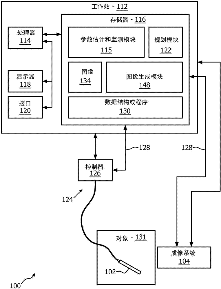
[0019] In accordance with the present principles, systems, methods, and apparatus are described for providing real-time feedback to a clinician or other operator during an ablation procedure. In particularly useful embodiments, the systems, methods and instruments employ shear wave elastography (SWE) as a modality for monitoring radiofrequency ablation (RFA) procedures. During RFA, the elastic properties of the tissue change so that the area being ablated becomes initially softer and progressively harder. Due to the dependence of shear wave velocity on underlying stiffness, SWE was able to discern the extent of lesion formation during RFA by determining the change in shear modulus during lesion formation. Different parameter sets for driving and tracking shear modulus or other shear wave or elastic parameters at various stages of the lesion formation process can be provided.

[0020] In accordance with the present principles, the systems, methods and instruments provide real-...


PUM


Abstract

A system for performing ablation includes an ablation device (102) configured to ablate tissue in accordance with control parameters and configured to make measurements during the ablation process. An imaging system (104) is configured to measure an elastographic related parameter to monitor ablation progress. A parameter estimation and monitoring module (115) is configured to receive the measurements from the ablation device and / or the elastographic related parameter to provide feedback to adaptively adjust imaging parameters of the imaging device at different times during an ablation process.

Description

technical field [0001] The present disclosure relates to ablation systems, methods and instruments, and more particularly to elastography measurements for optimizing ablation in medical applications. Background technique [0002] Thermal ablation techniques offer an excellent alternative to major surgery, which can pose risks to even the most experienced surgeon. These techniques are minimally invasive, requiring only needles (eg, radiofrequency (RF), cryotherapy, and microwave ablation) or noninvasive heat sources (such as using high-intensity focused ultrasound (HIFU)). In most procedures, cancerous tissue is heated above 60°C and coagulated. [0003] Radiofrequency ablation (RFA) is currently the only minimally invasive heat treatment approved by the FDA in the United States. It uses a probe with an active electrode tip through which a 460-500 kHz alternating current is conducted. The current travels through the body to ground pads that are placed on the patient's back...

Claims


Application Information

Patent Timeline
Patent Type & Authority Applications(China)
IPC IPC(8): A61B8/08A61B18/12
CPCA61B8/08A61B8/485A61B18/12A61B2018/00779A61B2018/00702A61B2018/00642A61B2018/00791A61B2018/00886A61B2090/378A61B34/10A61B34/25A61B18/00A61B2017/0019A61B2018/00577A61B2018/00684
Inventor S·巴拉特A·阿南德S·塞特拉曼黄圣文W·T·史
Owner KONINKLJIJKE PHILIPS NV
Why Eureka
Try Eureka