
Method for identifying, separating and culturing lymphoma stem cells from non-Hodgkin lymphoma cell line

A lymphoma cell, non-Hodgkin technology, applied in the field of cells, can solve the problem that cells cannot be equal to tumor stem cells, and achieve strong specific effects

Inactive Publication Date: 2017-11-17
QINGDAO UNIV
View PDF3 Cites 3 Cited by
  • Summary
  • Abstract
  • Description
  • Claims
  • Application Information

AI Technical Summary

Problems solved by technology

For a long period of time thereafter, due to the lack of research on the surface markers of lymphoma stem cells, the isolation and identification of lymphoma stem cells were often carried out through marginal cell (SP cell) isolation, limited dilution method, tumorigenicity detection, etc., but these isolation The method has a heavy speculative component, and the isolated cells cannot be equivalent to cancer stem cells
The isolation and identification methods of lymphoma stem cells need to be established urgently

Method used


Image

  • Method for identifying, separating and culturing lymphoma stem cells from non-Hodgkin lymphoma cell line
  • Method for identifying, separating and culturing lymphoma stem cells from non-Hodgkin lymphoma cell line
  • Method for identifying, separating and culturing lymphoma stem cells from non-Hodgkin lymphoma cell line

Examples


Embodiment 1

[0059] Example 1 Identification of Lymphoma Stem Cells from Non-Hodgkin's Lymphoma Cell Lines

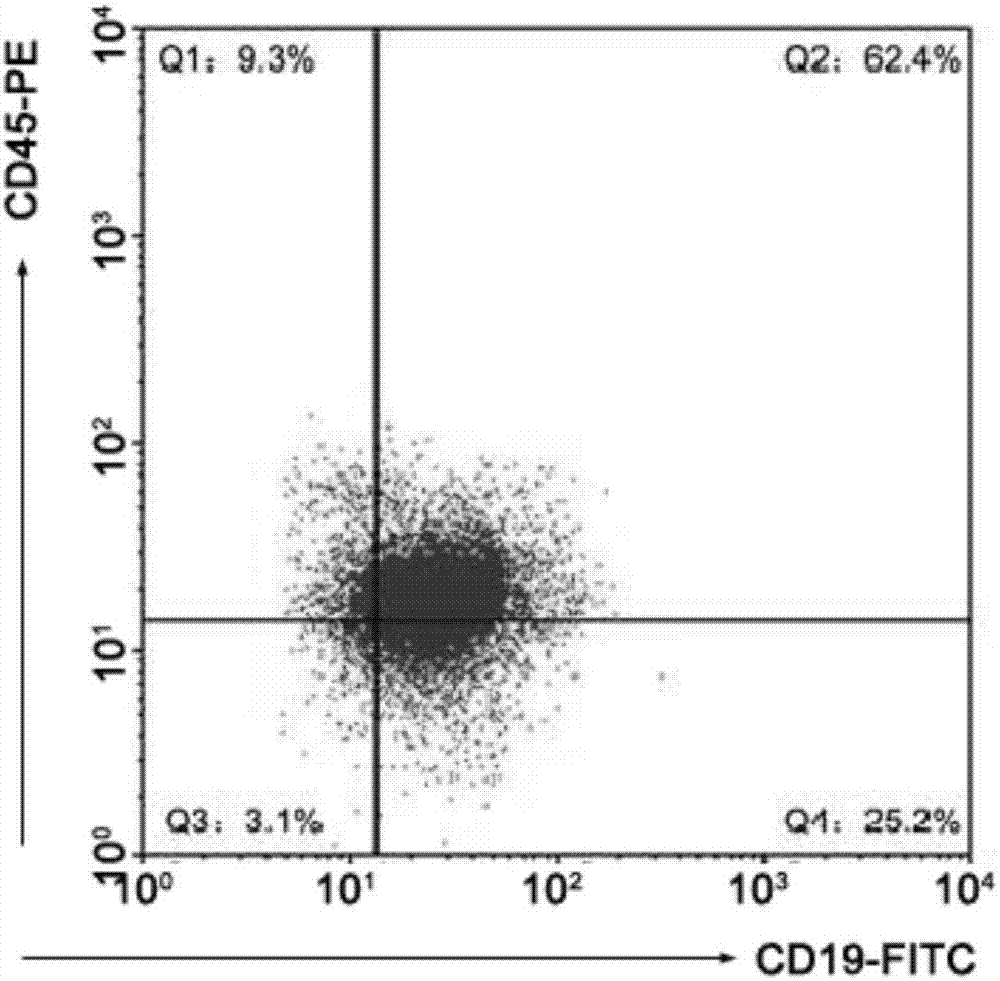
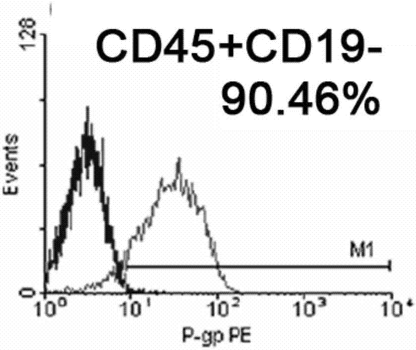
[0060] Identification of lymphoma stem cell content from non-Hodgkin's lymphoma cell lines, which may be CD45 + CD19 - cluster. Non-Hodgkin's lymphoma cells were routinely cultured in RPMI-1640 medium containing 10% fetal bovine serum. at 37°C, 5% CO 2 cultivated in an environment. Cells were passaged every three days and inoculated at a ratio of 1:4. After adjusting the cell state, the flow cytometric identification of CD45 and CD19 on the cell membrane surface was carried out. Adjust the number of cells to 2 × 10 5 cells / mL, add CD45-PE and CD19-FITC flow antibodies respectively, after dark staining for 30min, wash with PBS buffer three times. The cells were resuspended in PBS buffer, and the proportion of cells in each quadrant was measured by flow cytometry, and the total number of cells was 10,000. The ratio of CD marker in Raji cells is as follows figure 1 As shown, t...

Embodiment 2

[0061] Example 2 Isolation of Lymphoma Stem Cells from Non-Hodgkin's Lymphoma Cell Lines

[0062] Isolation of Lymphoma Stem Cells from Non-Hodgkin Lymphoma Cell Lines Using Magnetic Beads. Positive sorting (CD45 + ) and depletion sorting (CD45 + CD19 - ), and finally get CD45 + CD19 - cell subgroups.

[0063] Adjust the non-Hodgkin's lymphoma cells to 2 × 10 8 cells / mL, wash twice with pre-cooled PBS buffer, pass through a 70 μm sieve to remove cell clumps, centrifuge, and add 600 μL PBS buffer to resuspend. Add FCR blocking agent to the cell suspension to eliminate background staining, then add 200 μL CD45 Micro Beads-labeled immunomagnetic bead antibody, and incubate on ice for 45 minutes. Wash the cells twice with PBS, centrifuge to discard the supernatant, and add 500 μL PBS to resuspend. Put the separation column on the magnetic stand, add the cell suspension to the separation column, and let the CD45 + Cells adhere to the column and unlabeled cells flow out. A...

Embodiment 3

[0064] Example 3 Culture of Lymphoma Stem Cells Isolated from Non-Hodgkin's Lymphoma Cell Lines

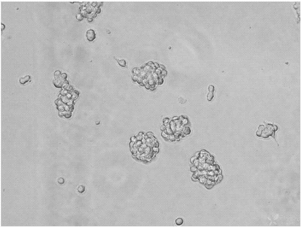
[0065] The non-Hodgkin's lymphoma stem cells provided in Example 2 of the present invention were cultured in a serum-free manner. The specific operation is: adjust the cell concentration to 500 cells / mL, and add 2 mL of cell suspension to each well of the six-well plate. Four growth factors, SCF, TPO, Flt-3L and IL-3, were added to each well so that the total concentrations were 100 μg / mL, 20 μg / mL, 50 μg / mL and 0.1 μg / mL, respectively. Place the six-well plate at 37 °C, 5% CO 2 Cultivate in an incubator, observe and take pictures after the formation of suspended cell spheres, this process takes about 1-2 weeks. figure 2 Shown is a suspension cell sphere formed by non-Hodgkin's lymphoma stem cells after two weeks of culture. It can be seen that the cells form a spherical clone cluster with clear boundaries and good refraction, indicating that the cells are all in a living state...


PUM


Abstract

The invention provides a method for identifying, separating and culturing lymphoma stem cells from a non-Hodgkin lymphoma cell line and relates to the technical field of cells. The method comprises the steps that surface markers of initiating cells of amphicyte lymphoma are identified and separated, and the specificity is high; meanwhile, the initiating cells of the amphicyte lymphoma express higher drug resistance to clinical frequently-used chemotherapy drugs, the lymphoma stem cells which are identified and separated by using the surface markers of the initiating cells are subjected to specific treatment, and the method has very important significance for preventing lymphoma relapse and improving the survival rate of patients. The method for identifying, separating and culturing the lymphoma stem cells from the non-Hodgkin lymphoma cell line can provide a research method and technical support for the mechanism research of non-Hodgkin lymphoma generation and development and the development of therapeutic drugs.

Description

technical field [0001] The invention relates to the field of cell technology, in particular to a method for identifying, separating and cultivating lymphoma stem cells from non-Hodgkin's lymphoma cell lines. Background technique [0002] Lymphoma is a malignant tumor originating from lymph nodes or extranodal lymphoid tissue. It usually grows as a solid tumor and is characterized by painless progressive lymphadenopathy, accompanied by symptoms such as weight loss and fever, and can involve other tissues and organs in the body. According to the data reported by the International Lymphoma Conference, there is a new case every 9 minutes in the world. In my country, there are 40,000 to 50,000 new cases every year, and more than 20,000 deaths. In the ranking of malignant tumor incidence, lymphoma accounts for It ranks 9th in male tumors, 10th in female tumors, and 3rd in childhood malignant tumors. The latest data show that Hodgkin's lymphoma accounts for about 8.54% of lymphoma ...

Claims


Application Information

Patent Timeline
IPC IPC(8): C12N5/095G01N33/574
CPCC12N5/0695C12N2501/125C12N2501/145C12N2501/2303C12N2501/26C12N2509/00G01N33/57484
Inventor 李培峰单婵王昆严考文
Owner QINGDAO UNIV
Learn More