
Image identification method and device

A recognition method and image technology, applied in the field of image processing, can solve the problems of occupying images, unsatisfactory diagnostic performance, cumbersome procedures, etc.

Inactive Publication Date: 2017-10-03
SHENZHEN UNIV
View PDF3 Cites 17 Cited by
  • Summary
  • Abstract
  • Description
  • Claims
  • Application Information

AI Technical Summary

Problems solved by technology

However, dermoscopy images may not always contain the entire lesion area, or the lesion area may only occupy a small portion of the image
In view of the large intra-class and inter-class differences between images of malignant skin lesions and benign skin lesions, the diagnostic performance provided by manually annotated features in existing methods is still unsatisfactory, especially the existing methods not only include complex and cumbersome procedures, and are laborious, time-consuming and subjective, resulting in poor generalizability and applicability in clinical practice

Method used


Image

  • Image identification method and device
  • Image identification method and device
  • Image identification method and device

Examples


no. 1 example

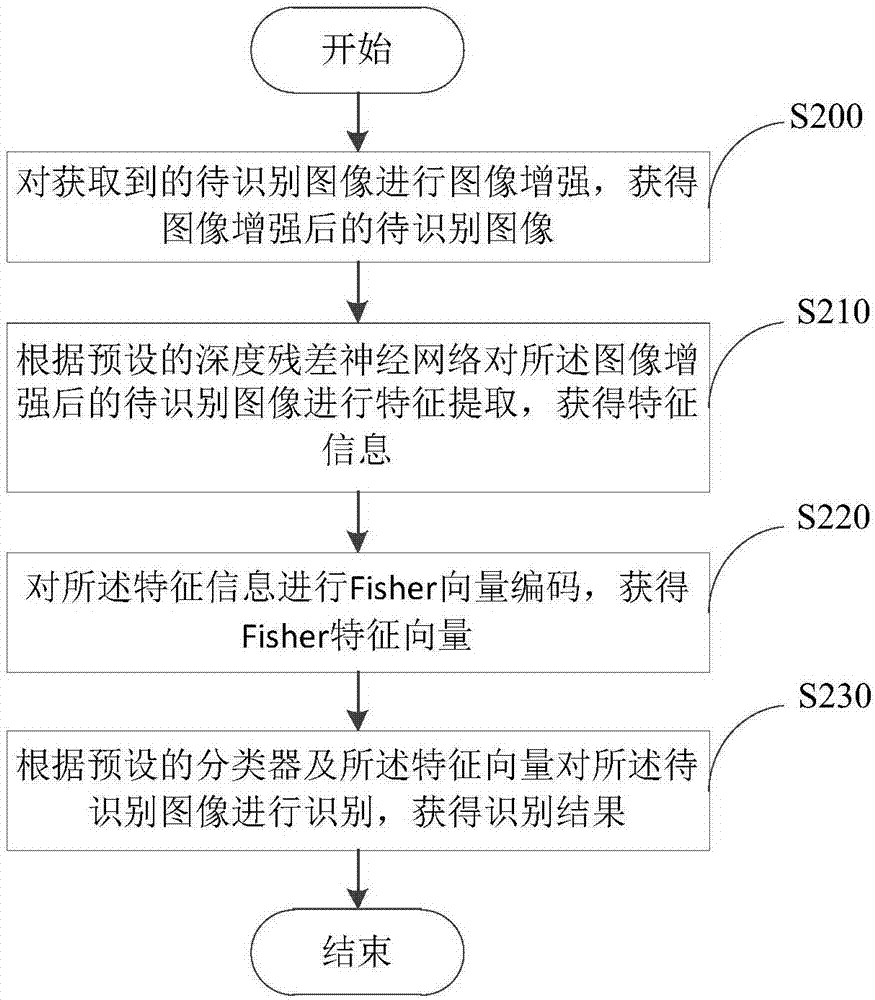
[0021] See figure 2 , An embodiment of the present invention provides an image recognition method, the method includes:

[0022] Step S200: Perform image enhancement on the acquired image to be recognized to obtain an image to be recognized after image enhancement.

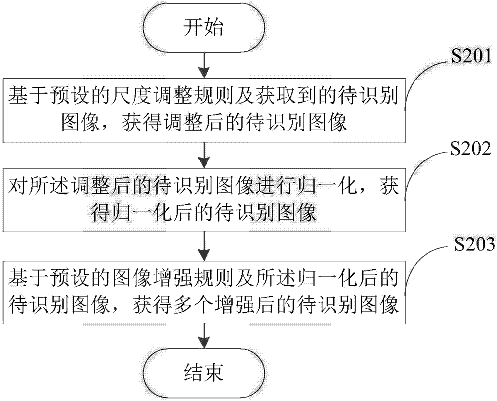
[0023] In order to ensure the accuracy of the features extracted from the image to be recognized, as an implementation, please refer to image 3 , The step S200 may include sub-step S201, sub-step S202, and sub-step S203.

[0024] Sub-step S201: Based on the preset scale adjustment rule and the acquired image to be recognized, an adjusted image to be recognized is obtained.

[0025] Since a deep residual neural network (Deep Residual Neural Network, ResNet) is used to extract features, a fixed and square size image is usually used as an input image, such as an image with a size of 227×227 or 224×224. Therefore, the image can be cropped and cropped to the required size for training or feature extraction. However, the res...

no. 2 example

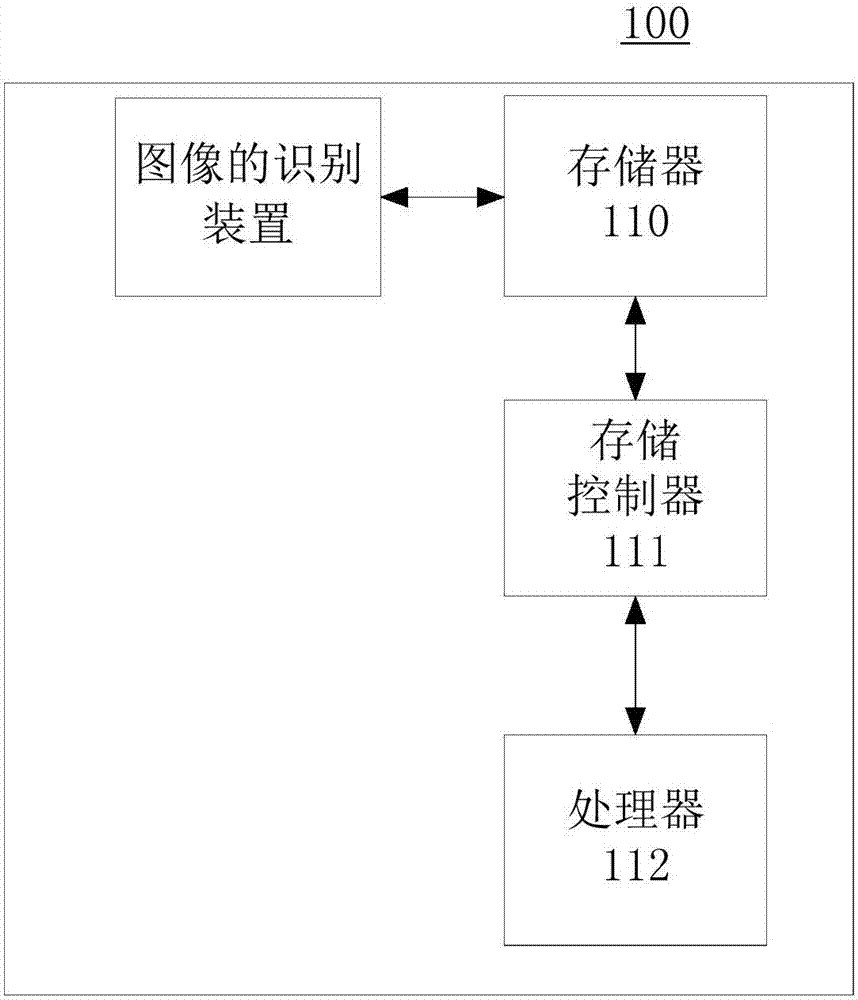
[0082] See Figure 4 An embodiment of the present invention provides an image recognition device 300, the device 300 includes: an image enhancement unit 310, a feature extraction unit 320, an encoding unit 330, and a recognition unit 340.

[0083] The image enhancement unit 310 is configured to perform image enhancement on the acquired image to be recognized to obtain the image to be recognized after the image enhancement.

Embodiment approach

[0084] As an implementation manner, the image enhancement unit 310 may include an adjustment subunit 311, a normalization subunit 313, and an image enhancement subunit 314.

[0085] The adjustment subunit 311 is configured to obtain the adjusted image to be recognized based on the preset scale adjustment rule and the acquired image to be recognized.

[0086] As an implementation manner, the adjustment subunit 311 may include an edge adjustment subunit 312.

[0087] The side adjustment subunit 312 is configured to adjust the short side of the acquired image to be recognized to a first preset value, and adjust the long side of the acquired image to be recognized to a second preset value, so that the adjustment The ratio of the short side and the long side of the subsequent image to be recognized is unchanged from the ratio of the short side and the long side of the acquired image to be recognized, so as to obtain the adjusted image to be recognized.

[0088] The normalization sub-unit 3...


PUM


Abstract

An embodiment of the invention discloses an image identification method and device and relates to the technical field of image processing. The method comprises the following steps: carrying out image enhancement on an obtained image to be identified to obtain an enhanced image to be identified; carrying out feature extraction on the enhanced image to be identified according to a preset deep residual neural network to obtain feature information; carrying out Fisher vector coding on the feature information to obtain a Fisher feature vector; and then, identifying the image to be identified according to a preset classifier and the feature vector to obtain an identification result. Therefore, more identification features of the image to be identified can be extracted by utilizing the deep residual neural network and Fisher vector coding; and then, the features are subjected to classification processing, and the image to be identified can be identified quickly according to the classification result. The method is simple to operate, is more efficient and more accurate, and is high in applicability.

Description

Technical field [0001] The present invention relates to the field of image processing technology, in particular, to an image recognition method and device. Background technique [0002] Melanoma skin cancer is one of the fastest growing and deadliest cancers in the world, accounting for 75% of skin cancer deaths. Early diagnosis is very important for the treatment of this disease because it can be easily cured in the early stages. In order to improve the diagnosis of this disease, dermoscopy is introduced to assist dermatologists in clinical examinations, because it is a non-invasive skin imaging technology that can provide clinicians with high-quality visual perception of skin damage. Compared with traditional macro (clinical) images, less surface reflections, deeper details and lower screening errors enable dermoscopy images to obtain better visibility and recognition accuracy. Since melanoma is more deadly than non-melanoma skin cancer, the difference between cancer and non-...

Claims


Application Information

Patent Timeline
Patent Type & Authority Applications(China)
IPC IPC(8): G06K9/62G06K9/46
CPCG06V10/44G06V10/462G06F18/2411
Inventor 雷柏英余镇汪天富倪东陈思平
Owner SHENZHEN UNIV
Learn More