
Quantitative analysis method suitable for abdominal subcutaneous fat of primate laboratory animal

A subcutaneous fat, experimental animal technology, applied in image analysis, image data processing, instruments, etc., can solve problems such as inability to obtain quantitative results, blurred MRI images, and indistinct distinctions.

Inactive Publication Date: 2017-06-13
广西南宁灵康赛诺科生物科技有限公司
View PDF9 Cites 4 Cited by
  • Summary
  • Abstract
  • Description
  • Claims
  • Application Information

AI Technical Summary

Problems solved by technology

However, the currently disclosed method is only applicable to the human body, and due to the characteristics of primate experimental animals that are different from the human body, such as small size and tight muscles, the obtained MRI images are often more blurred than those of the human body, and the distinction between regions is not obvious , so it is impossible to obtain accurate quantitative results when using existing processing and analysis technology solutions for quantitative analysis

Method used


Image

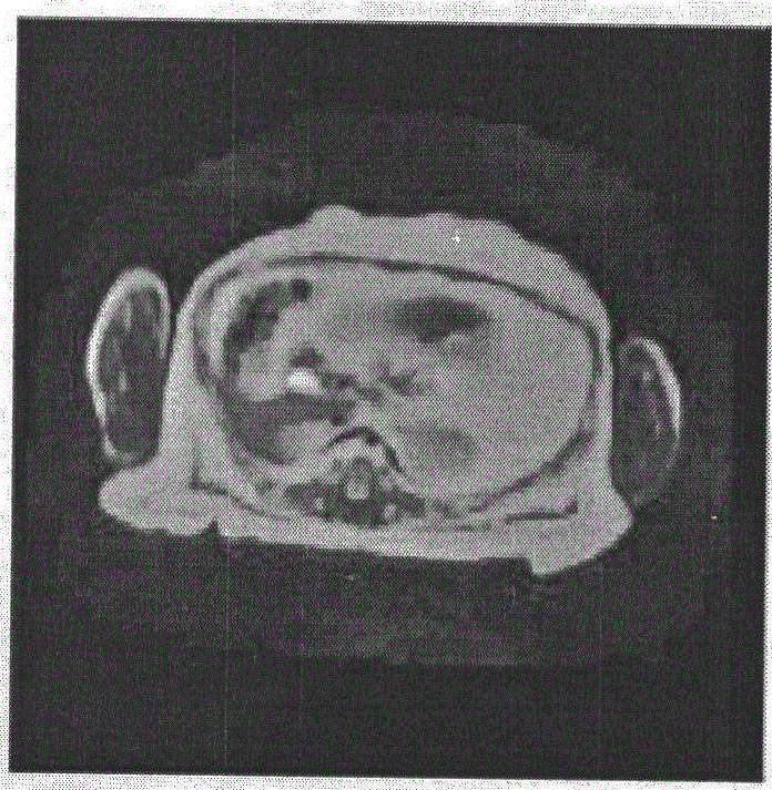
  • Quantitative analysis method suitable for abdominal subcutaneous fat of primate laboratory animal
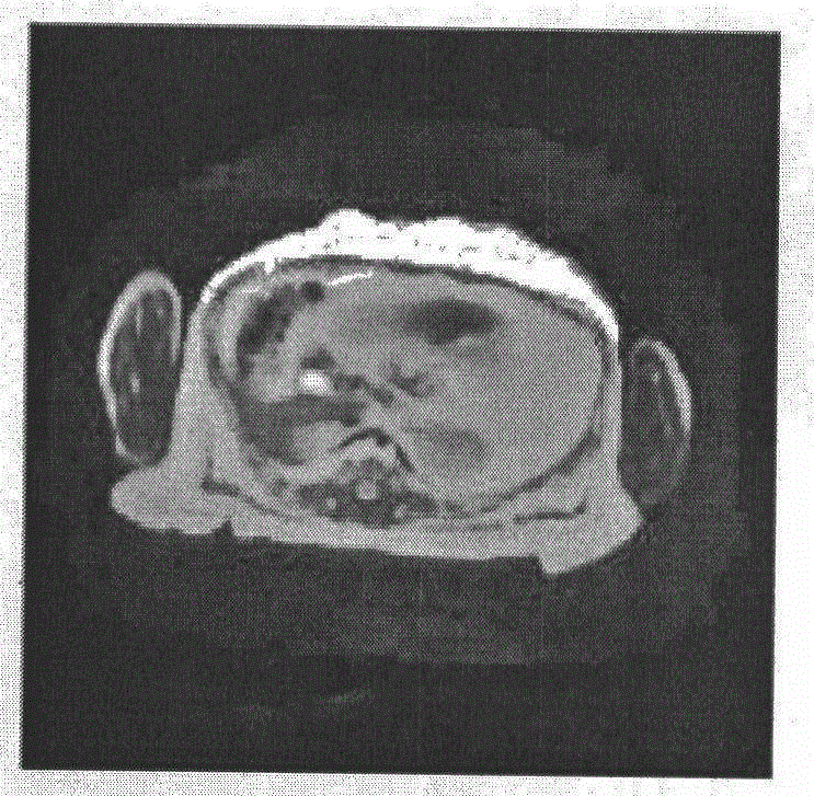
  • Quantitative analysis method suitable for abdominal subcutaneous fat of primate laboratory animal
  • Quantitative analysis method suitable for abdominal subcutaneous fat of primate laboratory animal

Examples


Embodiment 1

[0030] A method for quantitative analysis of abdominal subcutaneous fat suitable for primate experimental animals, comprising the steps of:

[0031] (1) The primate experimental animals are placed in a supine position. When the animals are placed, the center of the eyebrows, the tip of the nose, the middle gap of the incisors, the midpoint of the line connecting the nipples, and the navel of the animal are connected into a line, and the abdomen of the animal is scanned by MRI. After setting, the Dicom format T2 phase image and FS-FSE-T2WI lipid suppression image;

[0032] (2) Use MATLAB software to adjust the contrast enhancement of the above original image, use standardized histogram operation to equalize the image histogram, and use 6*6 median filter to denoise the median filter; use 9*9 Gaussian filter for Gaussian filter Filter denoising; the mean value filter is adjusted by using 9*9 mean value filter denoising image filtering, and the pixel value is stretched to 0-1;

...

Embodiment 2

[0039] A method for quantitative analysis of abdominal subcutaneous fat suitable for primate experimental animals, comprising the steps of:

[0040] (1) The primate experimental animals are placed in a supine position. When the animals are placed, the center of the eyebrows, the tip of the nose, the middle gap of the incisors, the midpoint of the line connecting the nipples, and the navel of the animal are connected into a line, and the abdomen of the animal is scanned by MRI. After setting, the Dicom format T2 phase image and FS-FSE-T2WI lipid suppression image;

[0041] (2) Use MATLAB software to adjust the contrast enhancement of the above original image, use standardized histogram operation to equalize the image histogram, and use 6*6 median filter to denoise the median filter; use 9*9 Gaussian filter for Gaussian filter Filter denoising; the mean value filter is adjusted by using 9*9 mean value filter denoising image filtering, and the pixel value is stretched to 0-1;

...

Embodiment 3

[0048] A method for quantitative analysis of abdominal subcutaneous fat suitable for primate experimental animals, comprising the steps of:

[0049] (1) The primate experimental animals are placed in a supine position. When the animals are placed, the center of the eyebrows, the tip of the nose, the middle gap of the incisors, the midpoint of the line connecting the nipples, and the navel of the animal are connected into a line, and the abdomen of the animal is scanned by MRI. After setting, the Dicom format T2 phase image and FS-FSE-T2WI lipid suppression image;

[0050] (2) Use MATLAB software to adjust the contrast enhancement of the above original image, use standardized histogram operation to equalize the image histogram, and use 6*6 median filter to denoise the median filter; use 9*9 Gaussian filter for Gaussian filter Filter denoising; the mean value filter is adjusted by using 9*9 mean value filter denoising image filtering, and the pixel value is stretched to 0-1;

...


PUM


Abstract

The invention discloses a quantitative analysis method suitable for abdominal subcutaneous fat of a primate laboratory animal. The method comprises the following steps of (1) allowing the primate laboratory animal to be in a lying manner, connecting a glabella, a nasal tip, a center joint between front teeth, a connection middle point between nipples and a navel of the animal into a line when the animal is put, and carrying out abdominal scanning on the animal by use of MRI (Magnetic Resonance Imaging); (2) carrying out contrast enhancement adjustment on an original image, equalizing an image histogram, adjusting image filtering, and extending a pixel value to be 0-1; (3) manually selecting a regional growth initial point by use of a regional growth method to complete image segmentation for the first time; (4) carrying out mean filtering again on the fat segmentation area for the first time completed in step (3), filtering, then fuzzifying an image region, and carrying out regional growth treatment for the second time; (5) calculating a cross sectional area of the corresponding abdominal subcutaneous fat in an ith-layer image by use of a formula Si=Ni*P; and (6) calculating a fat mass by use of the formula as shown in the description.

Description

technical field [0001] The invention relates to the technical field of quantitative analysis of abdominal subcutaneous fat of primate experimental animals under nuclear magnetic resonance images, in particular to a quantitative analysis method suitable for abdominal subcutaneous fat of primate experimental animals. Background technique [0002] At present, the detection means that people often adopt to obesity are: standard body weight method, body mass index (BMI) method, waist circumference and waist-to-hip ratio measurement method, sebum thickness method. However, the above-mentioned detection methods fail to involve the quantitative analysis of fat, and are only indirect detection methods, which have relatively large limitations. Although, there have been recent reports on quantitative analysis based on nuclear magnetic resonance images, such as the patent "Semi-automatic quantification method for human abdominal fat volume based on magnetic resonance images, application...

Claims


Application Information

Patent Timeline
Patent Type & Authority Applications(China)
IPC IPC(8): G06T5/00G06T7/11
CPCG06T2207/10088G06T2207/20032G06T2207/30088G06T5/94G06T5/92G06T5/70
Inventor 张国栋岳峰陶国现梁超李文
Owner 广西南宁灵康赛诺科生物科技有限公司
Learn More