Lung cancer targeted low-toxicity quantum dot preparation
A quantum dot and targeting technology, applied in the field of biotechnology and nano-biological materials, can solve the problems of high toxicity, difficult tumor tracing and detection, etc., and achieve the effect of simple preparation process and wide applicability
- Summary
- Abstract
- Description
- Claims
- Application Information
AI Technical Summary
Problems solved by technology
Method used
Image

Examples
Embodiment 1
[0033] Embodiment 1 prepares quantum dot preparation
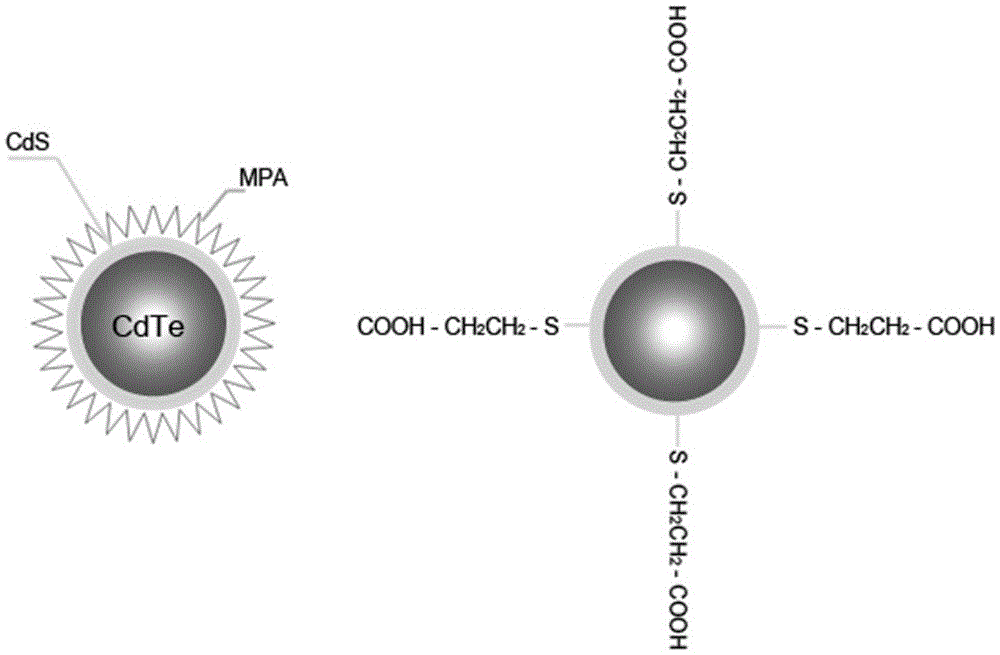
[0034] (1) Preparation of quantum dots: the appropriate amount of CdCl 2 , NaHTe and MPA are placed in the reactor, and NaBH with pH=9.0 4 After dissolving, react continuously at 95°C for 16 hours under the protection of nitrogen, and then add a certain amount of Na to the reactor 2 S, continuously reacted at 60°C for 1.5 hours, the ratio of Cd:Te:S:MPA in the reactor was 1:0.45:0.0125:4.161, and stored at 2°C;
[0035] (2) Preparation of quantum dots targeting lung cancer: Dissolve C-RGD in PBS (pH=7.4) to make a 1 mg / mL RGD short peptide solution (dissolved at 4°C), then molar ratio 1:1 Add the prepared quantum dots and react at 4°C for 4 hours;
[0036] (3) Preparation of quantum dot preparation: Dissolve chloroquine in 0.01M PBS (pH=7.4) and make a 10 μM chloroquine solution, then dissolve the prepared quantum dots with RGD in the solution so that the heavy metal concentration Cd 2+ = 1 μM; The identification of th...
Embodiment 2
[0039] Example 2. Comparison experiment of lung cancer targeting between quantum dots prepared in the present invention and ordinary quantum dots
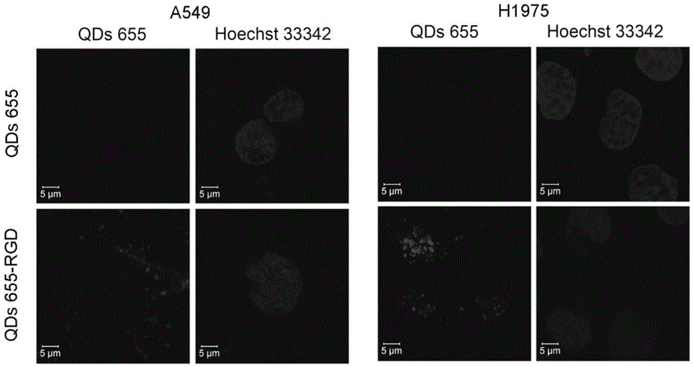
[0040] Lung cancer A549 cells and H1975 cells were seeded on laser confocal dishes (1×10 5 After 24 hours of incubation, the cells were allowed to adhere to the wall; after adding the quantum dots and common traditional quantum dots (20nM) and incubating for 2 hours, the cells were washed with PBS for 3 times and the situation of the cells bound to the quantum dots was observed by confocal laser, as shown in figure 2 As shown, A549 cells have no obvious fluorescence after co-incubating with traditional quantum dots for 2 hours, but there is obvious fluorescence in the cell membrane and cells after co-incubating with the quantum dot preparation targeting lung cancer for 2 hours, which proves that the quantum dots specifically target and Can bind to lung cancer cells.
Embodiment 3
[0041] Example 3. In vivo toxicity evaluation test of this quantum dot preparation
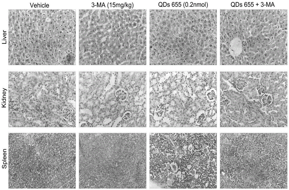
[0042] 0.2nmol of quantum dots and the prepared quantum dot preparations were injected into the mice through the tail vein respectively, and observed for 30 days respectively. No mice died in each group within 30 days. Anatomical analysis of its organs, the results are as follows image 3 As shown, after 30 days, the liver, kidney and spleen of common quantum dots were seriously damaged, and the damage caused by the prepared quantum dot preparation to the liver, kidney and spleen was significantly smaller than that caused by common quantum dots, proving that the prepared Quantum dot preparations have relatively low toxicity in vivo, and new preparations made with autophagy inhibitors can reduce the toxicity of quantum dots in vivo.
PUM
| Property | Measurement | Unit |
|---|---|---|
| particle diameter | aaaaa | aaaaa |
Abstract
Description
Claims
Application Information
 - R&D
- Intellectual Property
- Life Sciences
- Materials
- Tech Scout
- Unparalleled Data Quality
- Higher Quality Content
- 60% Fewer Hallucinations
Browse by: Latest US Patents, China's latest patents, Technical Efficacy Thesaurus, Application Domain, Technology Topic, Popular Technical Reports.
© 2025 PatSnap. All rights reserved.Legal|Privacy policy|Modern Slavery Act Transparency Statement|Sitemap|About US| Contact US: help@patsnap.com