OCT-based automatic detection and evaluation method and system for cardiovascular implanted stents

A coronary and image technology, applied in the field of automatic detection and evaluation of cardiovascular implanted stents based on OCT, can solve the problems of low intimal coverage, low stent accuracy, and high imaging quality requirements, and achieve high detection accuracy. , the effect of good robustness

Active Publication Date: 2020-04-10
SHENZHEN VIVOLIGHT MEDICAL DEVICE & TECH CO LTD
View PDF2 Cites 0 Cited by
  • Summary
  • Abstract
  • Description
  • Claims
  • Application Information

AI Technical Summary

Problems solved by technology

[0003] However, there are currently two problems in stent implantation and postoperative follow-up: 1. Due to the low resolution of X-ray contrast images, the situation of stent placement cannot be clearly known, resulting in poor stent placement during the operation, especially for stent placement. Second, due to poor stent apposition and other reasons, the degree of re-endothelialization of the postoperative stent is low, and the intima coverage is low, resulting in stent thrombosis, which seriously threatens the patient's life safety
The disadvantage of this method is that it is highly sensitive to noise such as blood artifacts, has high requirements for imaging quality, has low accuracy of stent detection, and does not have good robustness.

Method used


Image

  • OCT-based automatic detection and evaluation method and system for cardiovascular implanted stents
  • OCT-based automatic detection and evaluation method and system for cardiovascular implanted stents
  • OCT-based automatic detection and evaluation method and system for cardiovascular implanted stents

Examples


Embodiment Construction

[0046] Hereinafter, embodiments of the present invention will be described in detail with reference to the accompanying drawings. This invention may, however, be embodied in many different forms and should not be construed as limited to the specific embodiments set forth herein. Rather, the embodiments are provided to explain the principles of the invention and its practical application, thereby enabling others skilled in the art to understand the invention for various embodiments and with various modifications as are suited to particular intended uses.

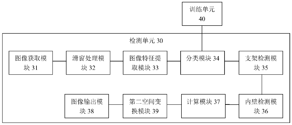
[0047] refer to figure 1 The OCT-based system for automatic detection and evaluation of cardiovascular implanted stents provided in this embodiment includes a detection unit 1 , an optical signal processing unit 2 , a data processing unit 3 and a display unit 5 connected in sequence. The detection unit 1 is used to collect the optical signal in the coronary artery, the optical signal processing unit 2 is used to process the ...


PUM


Abstract

The present invention provides a method and system for automatic detection and evaluation of an OCT-based cardiovascular implant stent, the method comprising the following steps: acquiring a coronary artery OCT image to be detected; Perform preprocessing; perform sliding window traversal on the preprocessed image; perform feature extraction on the sliding window image; judge whether the feature value of the extracted sliding window image is greater than the feature threshold corresponding to the classifier model; detect where the bracket is located in the sliding window image The area and the center of the stent; detect the inner wall contour of the coronary artery in the preprocessed image; calculate the shortest distance between the center of the stent and the inner wall contour of the coronary artery to obtain the adhering situation or coverage of the stent; the adhering situation of the stent or coverage is displayed. The method and system proposed by the invention are not affected by noise such as blood artifacts, can work in a complex imaging environment, have high detection accuracy of the stent, and have good robustness.

Description

technical field [0001] The invention relates to the technical field of medical devices, in particular to an OCT-based automatic detection and evaluation method and system for cardiovascular implanted stents. Background technique [0002] Coronary heart disease, also known as coronary atherosclerotic heart disease, is mainly caused by myocardial ischemia, hypoxia or necrosis caused by the accumulation of fatty substances and atherosclerotic plaques in the coronary artery wall. One of the diseases with the highest mortality rate. At present, the most important means of treating coronary heart disease is Percutaneous Coronary Intervention (PCI) surgery, which uses a special catheter with a balloon to be sent to the narrowed or blocked coronary artery through the peripheral artery. Inflate a balloon and place a stent to expand the lumen of the blood vessel, prevent restenosis of the blood vessel, and improve blood circulation in the blood vessel. [0003] However, there are cu...

Claims


Application Information

Patent Timeline
Patent Type & Authority Patents(China)
IPC IPC(8): G06T7/00G06T7/136G06K9/62A61B5/00
CPCA61B5/0066G06T7/0012A61B5/7235A61B5/7264G06T2207/20081G06T2207/10101G06F18/24133
Inventor 朱锐曹一挥
Owner SHENZHEN VIVOLIGHT MEDICAL DEVICE & TECH CO LTD
Why Eureka
Try Eureka