
Method for obtaining corneal endothelial cells

A corneal endothelium and acquisition method technology, which is applied in the field of obtaining corneal endothelial cells, and achieves the effects of simple technical solution, broad application prospect and important clinical significance.

Active Publication Date: 2020-05-19
EYECURE THERAPEUTICS INC JIANGSU
View PDF0 Cites 0 Cited by
  • Summary
  • Abstract
  • Description
  • Claims
  • Application Information

AI Technical Summary

Problems solved by technology

However, this therapy is significantly limited by the shortage of donor corneal endothelial cells

Method used


Image

  • Method for obtaining corneal endothelial cells
  • Method for obtaining corneal endothelial cells
  • Method for obtaining corneal endothelial cells

Examples


Embodiment 1

[0033] The configuration of embodiment 1 culture medium

[0034] 1. Preparation of serum-free medium

[0035] The basal medium is DMEM / F12 medium supplemented with 15% serum replacement, 15ng / mL basic fibroblast growth factor, 1× non-essential amino acid, 2mM GlutaMAX TM Medium and additives, 100 units / mL blue-chain double antibody, 0.1mM β-mercaptoethanol, prepared into a serum-free medium, stored at 4°C in the dark for future use.

[0036] 2. Preparation of differentiation medium A

[0037] The basal medium is DMEM / F12 medium supplemented with 1×N2 supplement, 12ng / mL basic fibroblast growth factor, 1×non-essential amino acid, 2mM GlutaMAX TM Culture medium and additives, 100 units / mL blue-chain double antibody, 2ng / mL recombinant human insulin, prepared as differentiation medium A, and stored at 4°C in the dark for future use.

[0038] 3. Preparation of Differentiation Medium B

[0039] The basal medium is DMEM / F12 medium supplemented with 10% serum replacement, 1× non-...

Embodiment 2

[0047] The acquisition method of embodiment 2 class corneal endothelial cells

[0048] 1. Cultivation of Commercialized Human Embryonic Stem Cells

[0049] Mouse embryonic fibroblasts were routinely cultured, and the 3-4 passages were taken as feeder layers, and the proliferation was induced in the medium supplemented with leukemia inhibitory factor (LIF), and the undifferentiated state was maintained.

[0050] 2. Acquisition of Embryoid Bodies

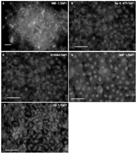
[0051] After the above-mentioned cultured commercial human embryonic stem cells were prepared and digested with collagenase IV and Dispase II at a ratio of 1:2, the prepared serum-free medium was used to culture human embryonic stem cells until they differentiated to form rosettes, that is, pseudo embryoid body, such as figure 1 as shown in a.

Embodiment 3

[0052] Embodiment 3 pre-differentiation culture

[0053] 1. Operation 1 of pre-differentiation culture

[0054] The embryoid bodies obtained in Example 2 were cultured in the differentiation medium A prepared in Example 1 supplemented with 500 ng / mL Noggin and 10 μM TGF-β1 receptor inhibitor for 4 days;

[0055] 2. Operation 2 of pre-differentiation culture

[0056] The embryoid bodies obtained in Example 2 were cultured in the differentiation medium B prepared in Example 1 supplemented with 500 ng / mL Noggin and 10 μM TGF-β1 receptor inhibitor for 2 weeks;


PUM


Abstract

The invention relates to an obtaining method of an analogous corneal endothelial cell, and belongs to the technical field of a cell biology. The analogous corneal endothelial cell is obtained through embryoid body obtaining, predifferentiation culture and differentiation culture. The analogous corneal endothelial cell obtained by a method of human multipotential stem cell in vitro differentiation can be used as the ideal cell resource for replacing the corneal endothelial cell for donation replacement. The method provided by the invention uses the commercial human embryonic stem cells; the commercial human embryonic stem cell has the self copying and unlimited proliferation capability and differentiation potential, so that the sufficient sources can be realized for the acquisition of the analogous corneal endothelial cell. The method provided by the invention solves the problem of low in vitro differentiation ratio of the analogous corneal endothelial cell; the in vitro differentiation ratio of the analogous corneal endothelial cell is improved; the time from the embryoid body stage to the analogous corneal endothelial cell obtaining is 11 days to the fastest degree. The method provided by the invention has the advantages that the technical scheme is simple and convenient; the operation is easy.

Description

technical field [0001] The invention relates to a method for obtaining corneal endothelial cells, belonging to the field of cell biology technology. Background technique [0002] Corneal endothelial cells are a monolayer of cells arranged in regular hexagons, which are the physiological barrier separating the corneal stroma from the aqueous humor and anterior chamber, and play a key role in maintaining corneal transparency. The reasons for the reduction or damage of corneal endothelial cells are: ① Gradually decrease with age; ② Infection, inflammation, trauma (such as mechanical injury, chemical injury, birth injury, laser rays, etc.), eye surgery (such as cataract surgery, anti-aging, etc.) Glaucoma surgery, and other intraocular operations (such as anterior chamber foreign body removal, etc.), genetic diseases, metabolic diseases, etc. can cause the reduction and damage of endothelial cells. Because adult corneal endothelial cells do not have the ability to proliferate, ...

Claims


Application Information

Patent Timeline
Patent Type & Authority Patents(China)
IPC IPC(8): C12N5/071C12N5/0735A61L27/38
CPCA61L27/3834A61L2430/16C12N5/0621C12N2500/38C12N2500/44C12N2500/90C12N2501/115C12N2501/135C12N2501/15C12N2501/33C12N2501/90C12N2501/998C12N2506/02
Inventor 范国平王云娟方攀峰李宁
Owner EYECURE THERAPEUTICS INC JIANGSU
Learn More