
A supporter for endoscopic submucosal tunnel

A supporter and tunnel technology, applied in the field of medical devices, can solve the problems of aggravating tunnel collapse, tunnel wall collapse, muscle layer damage, etc., and achieve the effect of improving resection efficiency and reducing perforation

Active Publication Date: 2019-05-07
LEO MEDICAL
View PDF6 Cites 0 Cited by
  • Summary
  • Abstract
  • Description
  • Claims
  • Application Information

AI Technical Summary

Problems solved by technology

The difficulty of this technique is that when the tunnel walls on both sides are finally resected, the tunnel walls collapse due to resection; at the same time, the superficial mucosa on the top of the tunnel shrinks, aggravating the collapse of the tunnel, resulting in a limited surgical field of view and affecting the resection efficiency. Some patients even Complications such as perforation caused by muscle layer injury

Method used


Image

  • A supporter for endoscopic submucosal tunnel
  • A supporter for endoscopic submucosal tunnel
  • A supporter for endoscopic submucosal tunnel

Examples


Embodiment Construction

[0028] The present invention will now be further described with reference to the accompanying drawings.

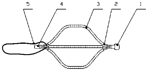
[0029] like Figure 1-6 As shown, an endoscopic submucosal tunnel supporter consists of a head 1, a skeleton 2, an insulating layer 3, a tail 4, a recovery line 5, an ejector rod 6, an outer sheath 7, an outer sheath handle 8, a jacking tube handle 9, etc. part composition. The head 1 , the skeleton 2 , the insulating layer 3 and the tail 4 constitute the support body 10 , and the support body 10 is shaped like a spherical, cylindrical, or diamond-shaped three-dimensional geometric body with an internal hollow shape. Several filamentary skeletons 2 are issued from the head 1 to the tail 4 and are arranged at a certain angle in the radial direction. The insulating layer 3 is wrapped around the skeleton 2 . The recycling line 5 is a thread, which passes through the wire gap of the skeleton 2 near the tail 4, and forms a closed-loop structure by knotting the head and tail ...


PUM


Abstract

The invention discloses an endoscopic submucosal tunnel support which is characterized by comprising a head, a framework, an insulating layer, a tail, a recycling wire, an ejector rod, a sheath, a sheath handle and an ejector rod handle. A support body comprises the head, the framework, the insulating layer and the tail; the recycling wire penetrates thread gaps, which are close to the tail, of the framework; a conveyor comprises the ejector rod, the sheath, the sheath handle and the ejector rod handle; the ejector rod is positioned in the sheath and can axially move; the support body is preliminarily compressed and gripped in the sheath and can be completely pulled away from the sheath after being released, and the original spatial stereoscopic geometric shape of the support body can be restored; the recycling wire can be hooked, dragged and pulled, so that the support body can be withdrawn. The endoscopic submucosal tunnel support has the advantages that tunnels can be effectively supported by the endoscopic submucosal tunnel support when tunnel walls are excised during endoscopic submucosal dissection, accordingly, complications such as perforation and inherent muscular layer injury can be reduced, and the excision efficiency can be improved.

Description

technical field [0001] The present invention relates to a medical device, in particular to a tunnel supporter for endoscopic submucosal dissection. Background technique [0002] Endoscopic submucosal dissection is an important technique for complete resection of early gastrointestinal and esophageal cancers through stomach and colonoscopy. The basic principle of the technique is to inject liquid into the submucosa through an injection needle under the guidance of a gastrointestinal endoscope to separate the mucosa from the muscular layer, and then gradually separate the submucosa through a variety of electrosurgical knives, so that the surface mucosa is completely peeled off from the muscular layer. . The tunneling technique is a commonly used technique in dissection. The method is to establish tunnel openings on the anal and oral sides of the lesion, and then gradually separate the submucosa with an electric knife from the oral side to the anal side, and finally connect w...

Claims


Application Information

Patent Timeline
Patent Type & Authority Patents(China)
IPC IPC(8): A61B17/02A61B17/00
CPCA61B17/00A61B17/0218
Inventor 刘岩闵敏黎洁张忠民
Owner LEO MEDICAL
Learn More