
A detection method for the biological activity of vascular endostatin

A technology of biological activity and vascular endothelium, which is applied in the field of detection of biological activity of vascular endostatin, can solve the problems of unstable quality, unstable method, poor sensitivity, etc., and achieve the effect of stable cell state and stable and reliable detection results

Active Publication Date: 2020-05-12
SHANDONG SIMCERE BIO PHARMA CO LTD
View PDF4 Cites 0 Cited by
  • Summary
  • Abstract
  • Description
  • Claims
  • Application Information

AI Technical Summary

Problems solved by technology

[0013] The disadvantages of the above method are: ①The ECGS used is the bovine pituitary gland extract, which is a mixture containing various endothelial cell growth factors, the extraction purity is low, and the quality is unstable, which leads to the instability of the method; ② This method is aimed at a variety of endothelial cell growth-promoting factors, which is not specific, and is interfered by a variety of non-specific cytokines, resulting in poor sensitivity of the method; ③The color development method used is MTT, which is cumbersome to operate

Method used


Image

  • A detection method for the biological activity of vascular endostatin
  • A detection method for the biological activity of vascular endostatin
  • A detection method for the biological activity of vascular endostatin

Examples


Embodiment 1

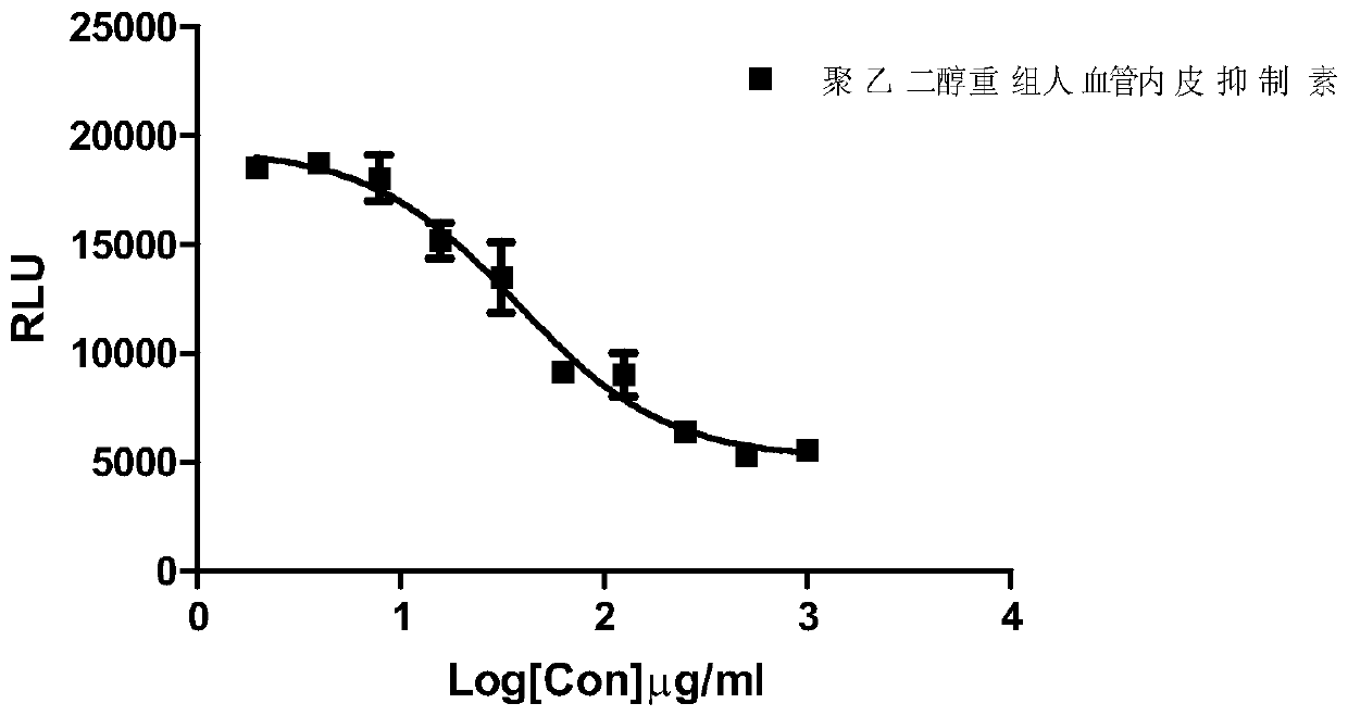
[0042] Example 1: PEGylated Recombinant Human Vascular Endostatin Inhibits bFGF Proliferation Activity of Human Umbilical Vein Endothelial Cells (HUVEC) Detection Method

[0043] The experimental steps are as follows:

[0044] (1) Cell culture and inoculation: After HUVEC cells (P3 generation, purchased from Sciencell) recovered, endothelial cell basal medium ECM (purchased from Sciencell), plus 5% FBS (purchased from Sciencell) and 1% ECGS (purchased from Sciencell) Sciencell, a mixture of various endothelial cell growth-promoting factors extracted from the bovine pituitary gland) was cultured at 37°C, 5% CO 2 In the incubator, replace the medium the next day; digest the cells with trypsin after reaching the logarithmic growth phase, centrifuge at 1200rpm for 5 minutes, discard the supernatant, and inoculate, and the remaining cells are passaged according to a certain proportion. Resuspend with starvation medium (ECM+1%FBS), and count live cells with a hemocytometer under a ...

Embodiment 2

[0064] Embodiment 2: polyethylene glycol recombinant human vascular endostatin inhibits the phosphorylation of bFGF on human umbilical vein endothelial cells (HUVEC) ERK

[0065] (1) HUVEC cell plating: use ECM (sciencell) + 5% FBS + 1% ECGS medium, according to 5 × 10 5 HUVEC cells were plated into 10 cm culture dishes (corning) at a density of cells / ml.

[0066] (2) Preparation of stimulating liquid: 1 ml of stimulating liquid was prepared as shown in Table 4, wherein bFGF was purchased from Shanghai Puxin Biotechnology Co., Ltd.

[0067] Table 4 Stimulation Solution Preparation Scheme

[0068]

[0069] (3) After 2 hours of starvation with ECM+0.5% FBS medium, the medium was aspirated, and washed twice with PBS equilibrated to room temperature, and then the stimulation solution was added, at 37°C, 5% CO 2 Next stimulate 8min. After the stimulation, add 5ml of ice-cold PBS to terminate the stimulation, place on ice, and use a cell scraper to collect the cells into a 15m...


PUM


Abstract

The invention discloses a method for measuring endostatin biological activity. Furthermore, the invention discloses a detection method of endostatin for inhibiting bFGF and promoting human umbilical vein vascular endothelial cell (HUVEC) proliferative activity. According to the method, endostatin obtained after double-dilution is premixed with a bFGF solution and then incubated with HUVEC cells simultaneously, a cell count staining reagent Cell titer-Glo is added, the chemiluminescence value is measured through an enzyme label reading meter, test data are processed with a computer program, and biological activity of an endostatin sample is calculated. The biological activity measured through the method is related to clinical effects and meets the technical requirement related to CFDA.

Description

technical field [0001] The invention relates to the technical field of biological product biological activity detection, and more specifically relates to a biological activity detection method of vascular endostatin. Background technique [0002] Vascular endostatin (Endostatin) is a kind of substance that O'Reilly isolated from the culture medium of the mouse endothelial cell line EOMAD in 1997 and has the effect of inhibiting vascular endothelial cells (O'Relly, M.S., etal.Cell.88: 277-285, 1997). Amino acid sequence analysis The substance is a degradation fragment of the carboxyl terminal of collagen XVIII, with a molecular weight of about 20kD. Endostatin can significantly inhibit the growth of various primary tumors in mice. Repeated use can make mouse tumors in a persistent dormant state, and will not develop drug resistance (Boehm, T. et al. Nature. 390: 404-407, 1997). [0003] Recombinant human endostatin (rhEndostatin) and recombinant mouse endostatin have 85% h...

Claims


Application Information

Patent Timeline
Patent Type & Authority Patents(China)
IPC IPC(8): G01N21/76
Inventor 熊新辉郑红霞仲恺
Owner SHANDONG SIMCERE BIO PHARMA CO LTD
Learn More