Medical imaging method and system
A medical imaging system and medical imaging technology, applied in image generation, image data processing, 2D image generation, etc., can solve problems such as poor operational flexibility and waste of resources, so as to improve efficiency, optimize user experience, and improve operational flexibility Effect
- Summary
- Abstract
- Description
- Claims
- Application Information
AI Technical Summary
Problems solved by technology
Method used
Image
Examples
Embodiment 1
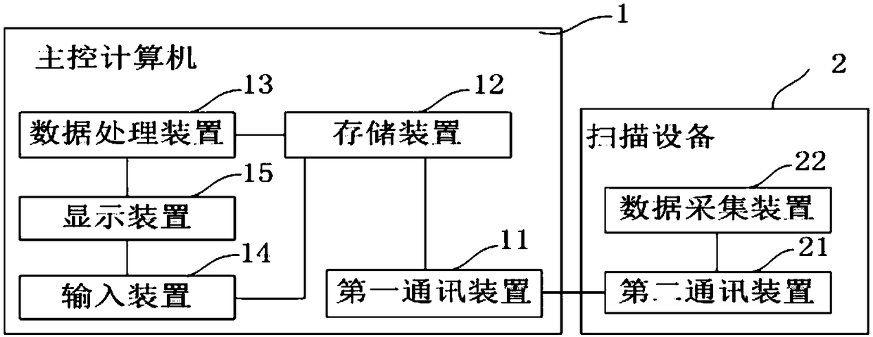
[0030] see figure 1 , the medical imaging system of the present invention includes a scanning device 2 and a main control computer 1, the scanning device 2 includes a second communication device 21 and a data acquisition device 22, and the main control computer 1 includes a first communication device 11, a storage device 12, and a data processing device. 13. Display device 15 and input device 14.
[0031] The display device 15 has a display interface, is provided with a data cutting mode option and a data cutting slider for selecting a data range, and the input device 14 is used for selecting the data cutting mode and setting the starting value and the starting value of data cutting for the original data. end value, and the start and end values of the data cut are displayed near opposite ends of the data cut slider. The storage device 12 is used for storing the original data and part of the original data corresponding to the start value and the end value of the data cutting...
Embodiment 2
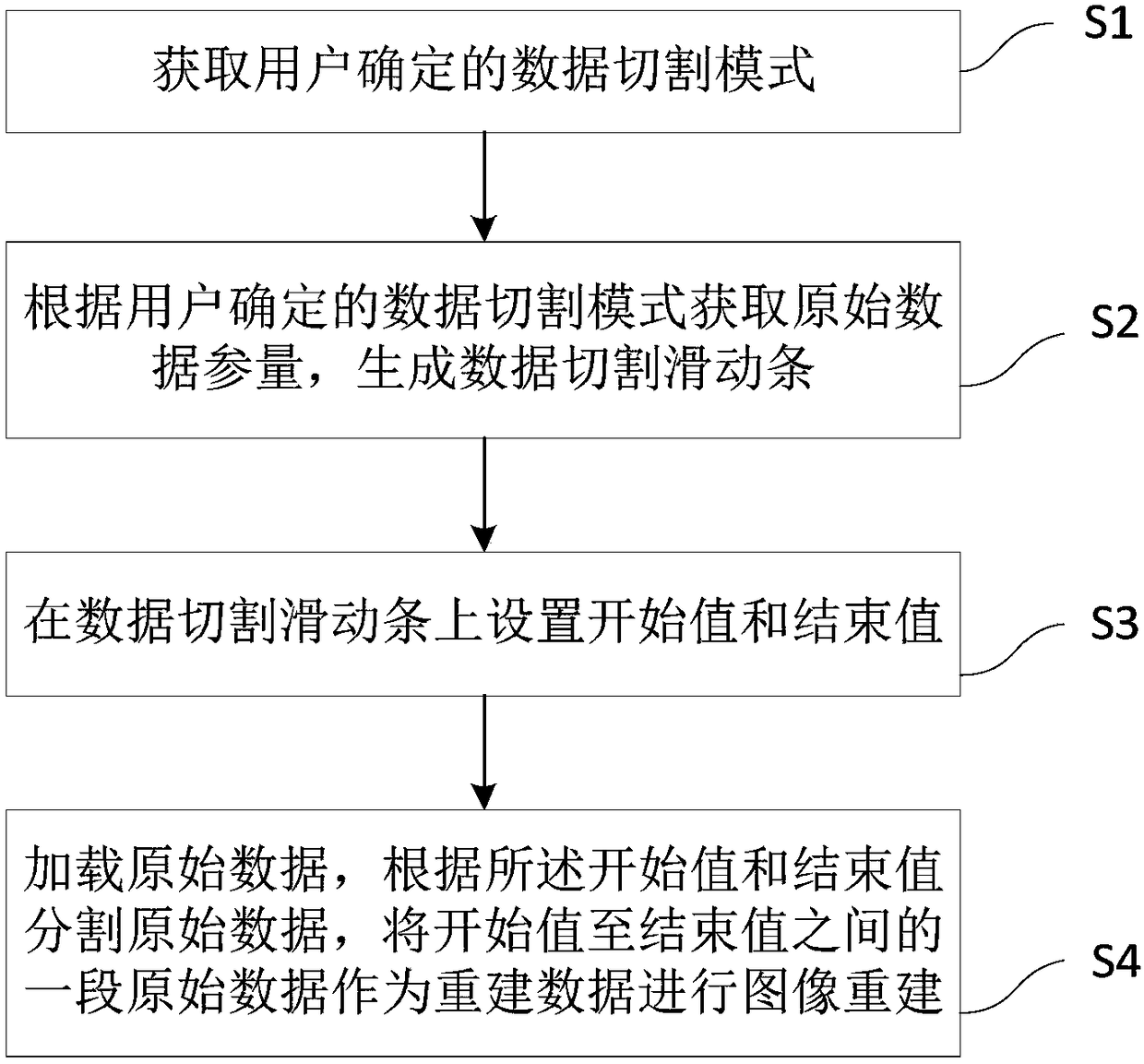
[0035] see figure 2 , the present invention provides a medical imaging method, the method is applied in the online scanning mode, including the following steps:
[0036] S1. Acquire a data cutting mode determined by a user, where the data cutting mode includes a time-based data cutting mode and a count-based data cutting mode.
[0037]Since the raw data acquisition is not performed or completed, only the time-based data cutting mode can be used in the online scan mode.
[0038] S2. Obtain the original data parameters according to the data cutting mode determined by the user, and generate a data cutting slider; in the online scanning mode, the original data parameters are the planned collection time of the original data.
[0039] S3. Set the start value and end value on the data cutting slider;
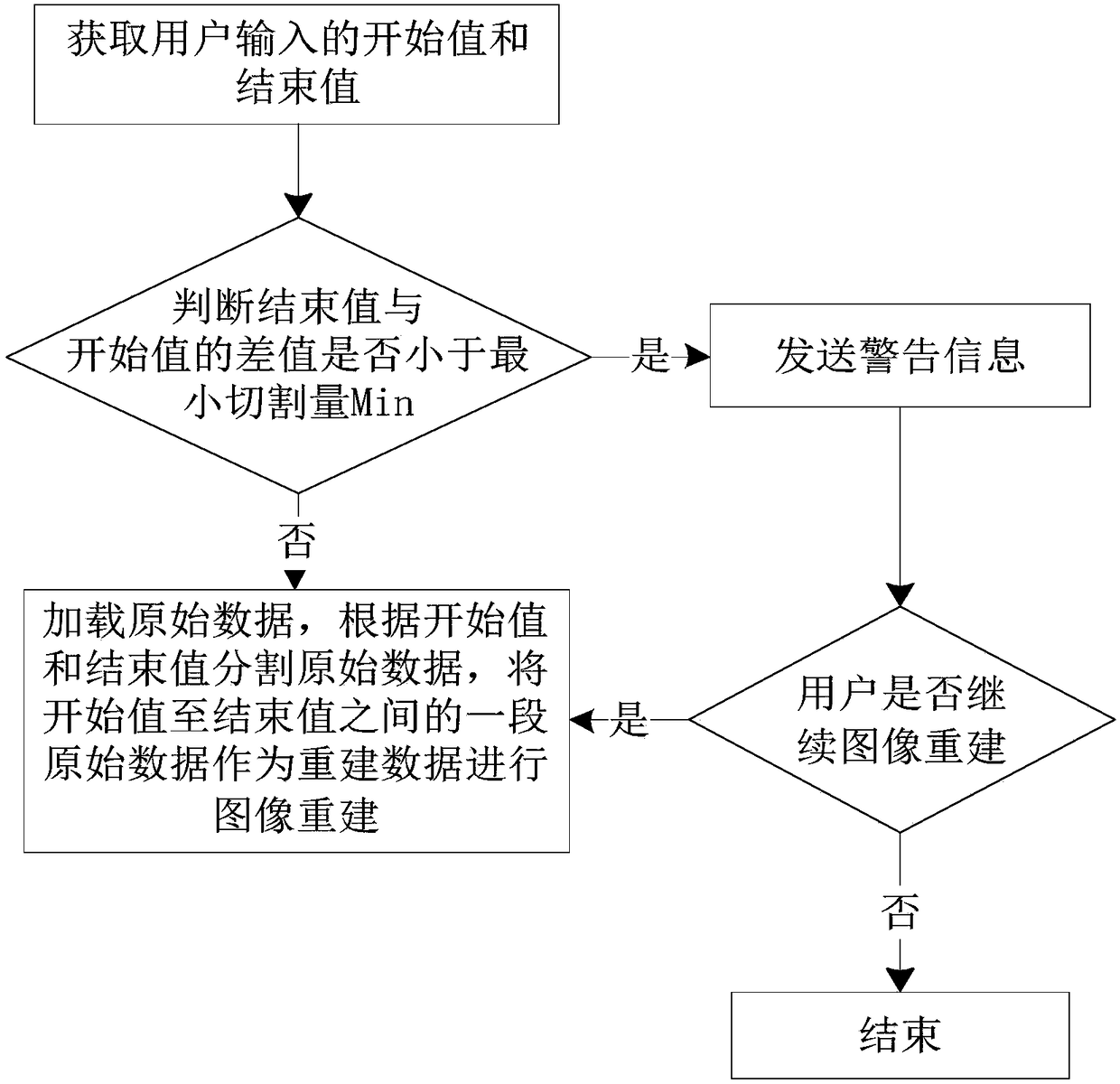
[0040] Specifically, the start value and the end value can be set by sliding the data to cut the cursor on the sliding bar or by directly inputting the value. On the data cutting s...
Embodiment 3
[0045] The present invention provides a medical imaging method, which is applied to an offline reconstruction mode, and includes the following steps:
[0046] S1. Acquire a data cutting mode determined by a user, where the data cutting mode includes a time-based data cutting mode and a count-based data cutting mode.
[0047] Since the acquisition of the original data in the offline reconstruction mode has been completed, and the acquisition time and amount of the original data have been obtained, in the offline reconstruction mode, both the time-based data cutting mode and the count-based data cutting mode can be selected for use.
[0048] S2. Obtain original data parameters according to the data cutting mode determined by the user, and generate a data cutting slide bar.
[0049] If the user selects the time-based data cutting mode, the raw data parameter is the collection time of the collected raw data; if the user selects the count-based data cutting mode, the raw data param...
PUM
 Abstract
Description
Claims
Application Information
 - R&D
- Intellectual Property
- Life Sciences
- Materials
- Tech Scout
- Unparalleled Data Quality
- Higher Quality Content
- 60% Fewer Hallucinations
Browse by: Latest US Patents, China's latest patents, Technical Efficacy Thesaurus, Application Domain, Technology Topic, Popular Technical Reports.
© 2025 PatSnap. All rights reserved.Legal|Privacy policy|Modern Slavery Act Transparency Statement|Sitemap|About US| Contact US: help@patsnap.com


