Animal jugular vein microdialysis method

A jugular vein and microdialysis technology, applied in blood sampling devices, medical science, sensors, etc., can solve problems such as few reports, and achieve the effect of less sampling, real and reliable sampling data, and avoiding stress

Inactive Publication Date: 2016-05-25
WUXI APPTEC SUZHOU +2
View PDF3 Cites 2 Cited by
  • Summary
  • Abstract
  • Description
  • Claims
  • Application Information

AI Technical Summary

Problems solved by technology

At present, microdialysis technology has become one of the important research tools of experimental neurophysiology and neurochemistry, which can provi

Method used


Image

  • Animal jugular vein microdialysis method

Examples


Embodiment 1

[0020] Embodiment 1 rat jugular vein microdialysis method

[0021] 1. Experimental materials:

[0022] Anesthetics: isoflurane (or 20% pentobarbital sodium);

[0023] Surgical instruments: ophthalmic scissors, ophthalmic forceps, glass minute needles, arterial clips, puncture needles (used to penetrate animal skin), iodophors, 75% alcohol cotton balls, dry cotton balls, sterile gloves, sterile gauze, etc.;

[0024] Jugular vein microdialysis probe: MD-2305 or MD-2310BASi;

[0025] Awake animal free movement device: Reman AIS;

[0026] Injection pump: AS50 automatic syringe pump;

[0027] Experimental animals: 300-400g SD rats.

[0028] 2. Experimental steps

[0029] Step 1. Rats are induced anesthetized with 4%-5% isoflurane and maintained anesthesia with 2%-3% isoflurane. During the operation, the anesthetized state of the animal should be monitored and the body temperature of the animal should be maintained.

[0030] Step 2: After the animal is under anesthesia, plac...


PUM


Abstract

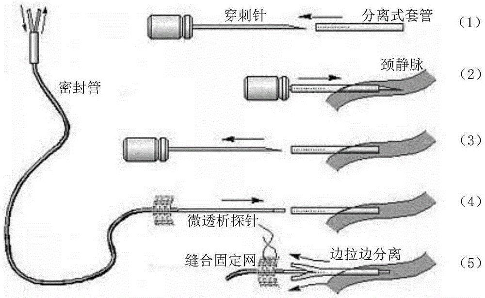
The invention discloses an animal jugular vein microdialysis method. The method comprises the steps that 1, an animal is anesthetized, hair on the neck is shaved, the neck is disinfected, and one side of the neck is vertically cut open; 2, fat and connective tissue around the jugular vein are subjected to blunt dissection; 3, under the skin of the neck and back on the surgical side, the subcutaneous bursa cavity is subjected to blunt dissection, and a channel is pierced out; 4, a syringe needle of a separate trocar is used for piercing the blood vessel at the end, away from the heart, of the jugular vein, a cannula is pushed into the jugular vein, the syringe needle is pulled out, a microdialysis probe is inserted in the jugular vein through the cannula and fixed, and the cannula is taken out; 5, a catheter of the microdialysis probe is led out of the body, and an injection pump is connected to the catheter for injection; 6, after injection is over, the incision is sutured. According to the method, microdialysis is performed through a jugular vein intubation method, the advantages of a small sampling quantity, little tissue damage and more truthful and reliable data are achieved, continuous sampling and real-time monitoring can be performed on a sober animal which moves freely, quantitative analysis can be carried out, and the method can be used as an effective means for drug research on in-vivo or in-vitro compounds.

Description

technical field [0001] The invention relates to the biological field, in particular to an animal jugular vein microdialysis method. Background technique [0002] Information such as the distribution and concentration of endogenous and exogenous compounds in the body is generally analyzed by collecting blood or tissue homogenate. With the deepening of research, this conventional method gradually shows many limitations. For example, for endogenous compounds, plasma or tissue homogenate can only provide basic information such as concentration, but the release mode and dynamic balance of the compound in the body cannot be reflected. [0003] Microdialysis (Microdialysis) technology is a new technology that combines perfusion sampling and dialysis technology and gradually improves dynamic micro-biological sampling from living organisms. It has continuous sampling, dynamic observation, quantitative analysis, small sampling volume, It has the advantages of light tissue damage, ca...

Claims


Application Information

Patent Timeline
IPC IPC(8): A61B5/15A61B5/155A61B5/153
CPCA61B5/150992A61B5/153A61B5/155
Inventor 薛彬金毅周聪颖王福强张伟军胡璇毕玲玲谢瑞东王新峰赵莹
Owner WUXI APPTEC SUZHOU
Why Eureka
Try Eureka