A preparation method of fluorescence-magnetic resonance dual-mode carbon quantum dots
A carbon quantum dot and magnetic resonance technology, applied in the fields of chemistry and biomedicine, can solve the problems of inability to detect tissues and organs, insufficient depth of tissue imaging, and inability to apply clinical disease detection, etc. The effect of shortening the relaxation time
- Summary
- Abstract
- Description
- Claims
- Application Information
AI Technical Summary
Problems solved by technology
Method used
Image
Examples
preparation example Construction
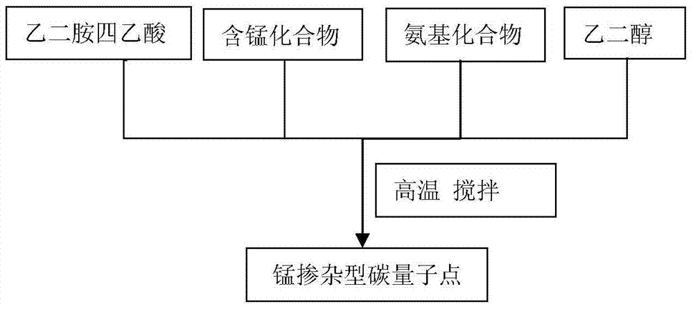
[0026] Such as figure 1 As shown, a method for preparing a fluorescent-magnetic resonance dual-mode nanoprobe of the present invention comprises the following methods:
[0027] (1) Dissolve ethylenediaminetetraacetic acid and manganese-containing compounds with a molar ratio of 1:0.05 to 1:2 in an amino compound and ethylene glycol solvent with a volume of 1:10 to 10:1. Magnetic stirring is carried out under high temperature conditions, and the reaction time is 4h-48h to obtain a dark brown homogeneous solution;
[0028] (2) Put the solution prepared in step (1) into a dialysis bag (MWCO: 1000Da) for dialysis, the dialysis time is 48h~72h, and change the water every 6h;
[0029] (3) The dialysis product in step (2) is subjected to vacuum rotary evaporation to a solid state, the evaporation temperature is 50 ° C, and the pressure is -0.1 MPa, that is, manganese-doped carbon quantum dots are obtained, which are recorded as Mn-CQDs;
[0030] The particle size of the manganese-d...
Embodiment 1
[0033] Dissolve 0.5mmol ethylenediaminetetraacetic acid and 1.0mmol manganese chloride (molar ratio 1:2) into 2mL triethylenetetramine and 5mL ethylene glycol, stir magnetically at a high temperature of 200°C, and the reaction time is 4h. Obtain a dark brown homogeneous solution; put the gained solution into a dialysis bag (MWCO: 1000Da) for dialysis, the dialysis time is 72h, and change the water every 6h; the product after the dialysis is vacuum rotary evaporated to a solid state, the evaporation temperature at 50°C and a pressure of -0.1MPa to obtain manganese-doped carbon quantum dots, denoted as Mn-CQDs;
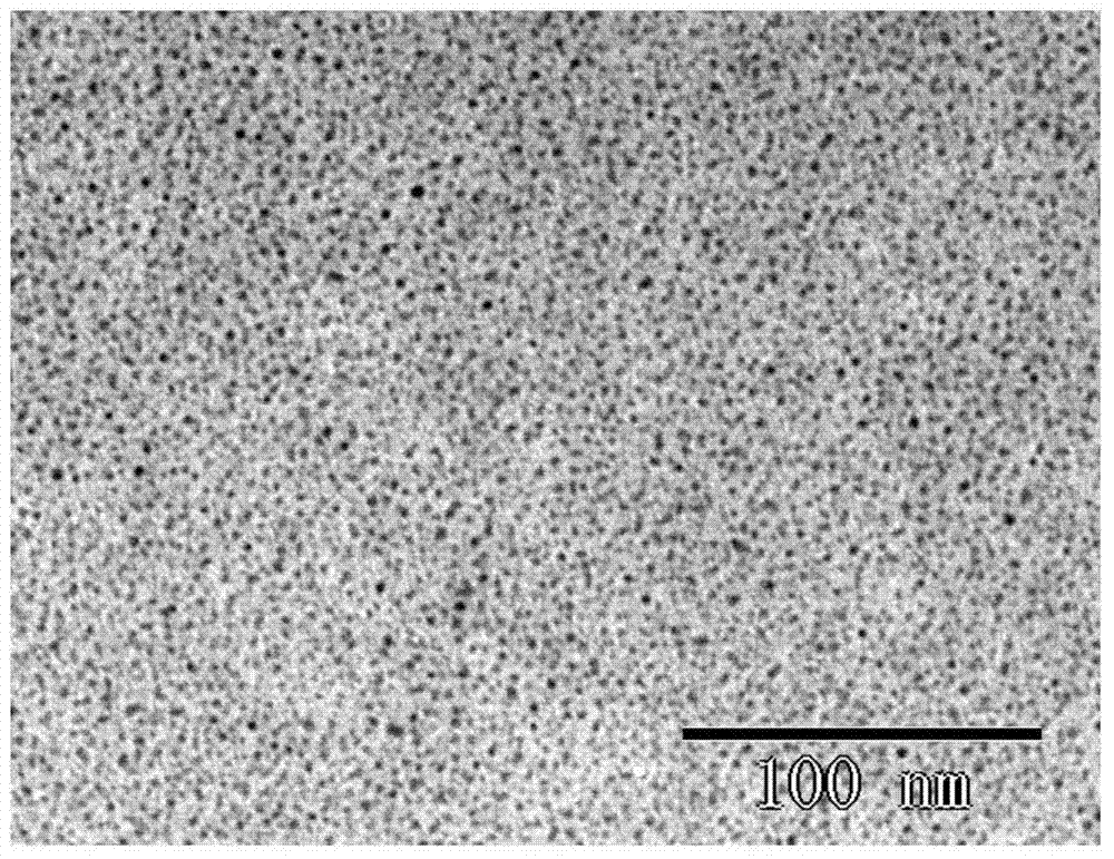
[0034] The particle size of the manganese-doped carbon quantum dot is 1.2nm-4.9nm, and it emits bright blue fluorescence under an ultraviolet lamp (365nm), its fluorescence emission peak is at 450nm, and its relaxation efficiency is 3.07±0.26mM -1 the s -1 .
Embodiment 2
[0036] Take 0.5mmol of ethylenediaminetetraacetic acid and 1.0mmol of manganese chloride (molar ratio 1:2) into 2mL of triethylenetetramine and 5mL of ethylene glycol, and stir magnetically at 120°C for 26h to obtain dark brown uniform quality solution; put the obtained solution into a dialysis bag (MWCO: 1000Da) for dialysis, the dialysis time is 60h, and change the water every 6h; the product after dialysis is vacuum rotary evaporated to a solid state, and the evaporation temperature is 50°C. Manganese-doped carbon quantum dots were obtained at a pressure of -0.1MPa, denoted as Mn-CQDs;
[0037] The particle size of the manganese-doped carbon quantum dots is 0.7nm-4.9nm, and it emits bright blue fluorescence under an ultraviolet lamp (365nm), its fluorescence emission peak is at 436nm, and its relaxation efficiency is 2.97±0.07mM -1 the s -1 .
PUM
| Property | Measurement | Unit |
|---|---|---|
| diameter | aaaaa | aaaaa |
| particle diameter | aaaaa | aaaaa |
| particle diameter | aaaaa | aaaaa |
Abstract
Description
Claims
Application Information
 - R&D
- Intellectual Property
- Life Sciences
- Materials
- Tech Scout
- Unparalleled Data Quality
- Higher Quality Content
- 60% Fewer Hallucinations
Browse by: Latest US Patents, China's latest patents, Technical Efficacy Thesaurus, Application Domain, Technology Topic, Popular Technical Reports.
© 2025 PatSnap. All rights reserved.Legal|Privacy policy|Modern Slavery Act Transparency Statement|Sitemap|About US| Contact US: help@patsnap.com


