
Tri-modal breast imaging system and imaging method thereof

A breast imaging and imaging system technology, applied in the fields of ultrasound/sonic/infrasonic Permian technology, ultrasound/sonic/infrasonic image/data processing, medical science, etc., can solve the problems of unfavorable disease diagnosis and classification, and reduce tumors Misdiagnosis rate, improved specificity and sensitivity, high contrast effect

Inactive Publication Date: 2015-08-12
PEKING UNIV
View PDF11 Cites 11 Cited by
  • Summary
  • Abstract
  • Description
  • Claims
  • Application Information

AI Technical Summary

Problems solved by technology

At present, photoacoustic tomography has not been widely used clinically, and its use alone is not conducive to the diagnosis and classification of diseases

Method used


Image

  • Tri-modal breast imaging system and imaging method thereof
  • Tri-modal breast imaging system and imaging method thereof
  • Tri-modal breast imaging system and imaging method thereof

Examples


Embodiment 1

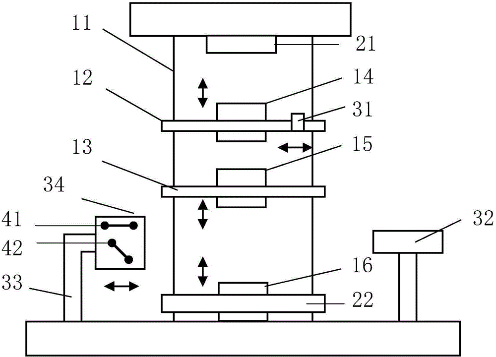
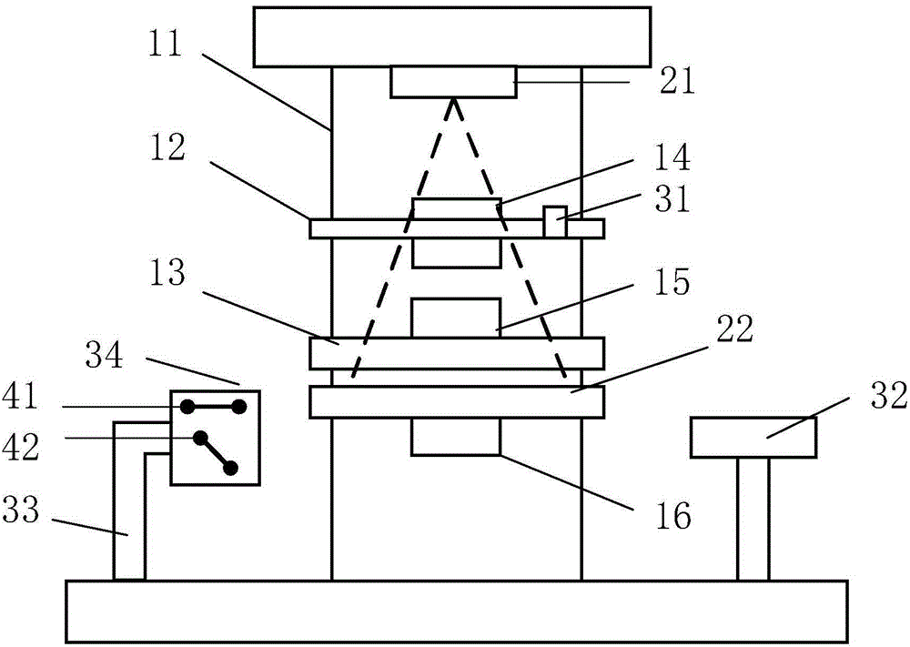
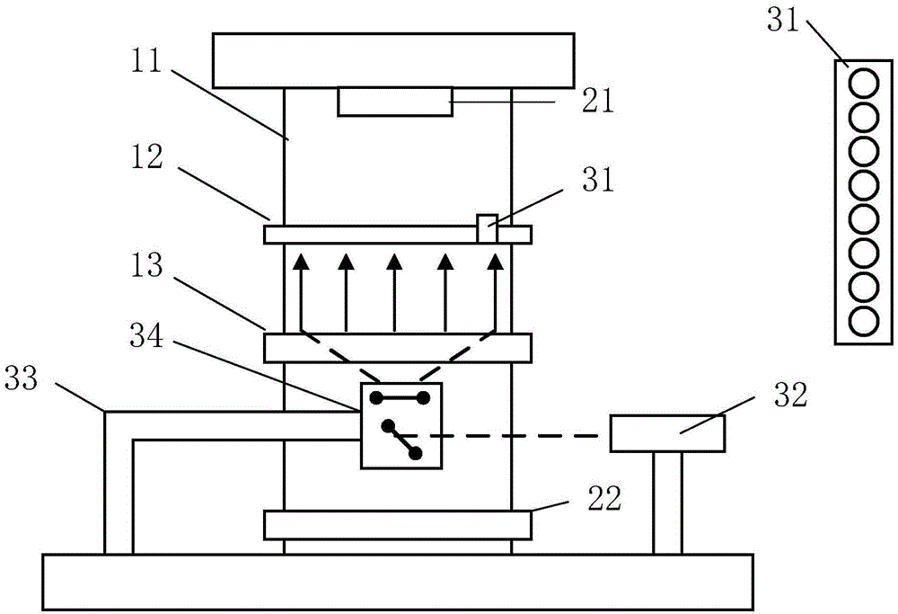
[0045] Such as figure 1 As shown, in this embodiment, the refractive diverging element 34 is arranged on the translation platform. The three-mode breast imaging system of the present embodiment includes: a frame 11, a pressing plate 12, a transparent plate 13, a first lifting platform 14, a second Lifting platform 15, third lifting platform 16, X-ray source 21, X-ray detector 22, ultrasonic array 31, laser 32, moving platform 33, refraction divergence element 34, and concave lens group 41 and plane mirror 42 constituting the refraction divergence element; Wherein, the lifting platform includes a track and a platform, the track is fixed on the frame, the direction of the track is the axis of the lifting platform, the platform is arranged in the track, and can move along the track; the frame 11 is placed vertically, and the first to the third lifting platform Vertically fixed on the frame, the pressing plate 12 and the transparent plate 13 are respectively fixed on the platforms...

Embodiment 2

[0054] Such as Figure 5 As shown, in this embodiment, the X-ray detector 22 and the refraction diverging element 34 are arranged on the conversion table 17. The three-mode breast imaging system of the present embodiment includes: a frame 11, a pressing plate 12, a transparent plate 13, a second A lifting platform 14, a second lifting platform 15, a third lifting platform 16, an X-ray source 21, an X-ray detector 22, an ultrasonic array 31, a refractive diverging element 34, a laser 32 and a computer; , the pressing plate 12 and the transparent plate 13 are respectively fixed on the first and second lifting platforms 14 and 15; the tissue to be measured is placed between the pressing plate 12 and the transparent plate 13 and fixed; along the axis of the lifting platform, it is arranged on the outside of the pressing plate 12 The X-ray source 21 is provided with a conversion table 17 on the outside of the transparent plate 13; the X-ray detector 22 and the refraction divergence...


PUM


Abstract

The invention discloses a tri-modal breast imaging system and an imaging method thereof.A testing system of the tri-modal breast imaging system comprises a frame, first, second and third lifting platforms, a pressing plate, a transparent plate, an X-ray source, an X-ray detector, an ultrasonic array, a laser, a data collection card, a computer, a refractive divergence component and a motion platform or a transfer platform. By means of integration among three modals including photoacoustic tomography, X-ray breast imaging and ultrasonic imaging, one piece of imaging equipment is adopted for tri-modal breast imaging without changing positions of tissue to be tested. The three modals differ in imaging contrast sources and complement with one another in terms of information. Therefore, non-invasive imaging among the structure, physiological and pathological features and the metabolic function of mammary tissue is carried out after images are filtered, projected and fused so that position and size of a tumor can be accurately determined. The tri-modal breast imaging system is particularly suitable for early detection and continuous monitoring of treatment effect of a breast cancer.

Description

technical field [0001] The invention relates to breast imaging technology, in particular to a three-mode breast imaging system including photoacoustic tomography, ultrasonic imaging and X-ray breast imaging and an imaging method thereof. Background technique [0002] According to the statistics of the World Health Organization, 1.2 million women in the world suffer from breast cancer every year, and 500,000 women die of breast cancer. The incidence rate is increasing at a rate of 2% to 8% every year. Approximately 100 million women are currently screened for breast cancer each year. Ultrasound imaging (Ultrasound) and X-ray mammography (Mammography) have been widely used in early breast cancer screening. Although ultrasonography has the advantages of non-destructive, non-invasive, portable, and good distinction between solid tumors and cystic lesions, the sensitivity and specificity of ultrasonography are insufficient, and it is not sensitive to imaging of tiny calcificatio...

Claims


Application Information

Patent Timeline
IPC IPC(8): A61B6/00A61B8/08A61B5/00
Inventor 李长辉丁翼晨任秋实
Owner PEKING UNIV
Learn More