
Vein imaging method for visible-light skin images

A skin image and vein imaging technology, which is applied in the directions of using light for diagnosis, medical science, instruments, etc., can solve the problems of inability to directly observe, unable to process and detect vein images, and achieve the effect of simple processing.

Active Publication Date: 2015-06-10
NANJING UNIV OF AERONAUTICS & ASTRONAUTICS
View PDF5 Cites 17 Cited by
  • Summary
  • Abstract
  • Description
  • Claims
  • Application Information

AI Technical Summary

Problems solved by technology

Usually, capillaries are located on the surface of the skin and can be observed with the naked eye, while veins are located below the dermis of the human skin, which cannot be directly observed from visible light images, making this method unable to process and detect vein images

Method used


Image

  • Vein imaging method for visible-light skin images
  • Vein imaging method for visible-light skin images
  • Vein imaging method for visible-light skin images

Examples


Embodiment Construction

[0044] The present invention will be described in further detail below in conjunction with specific embodiments.

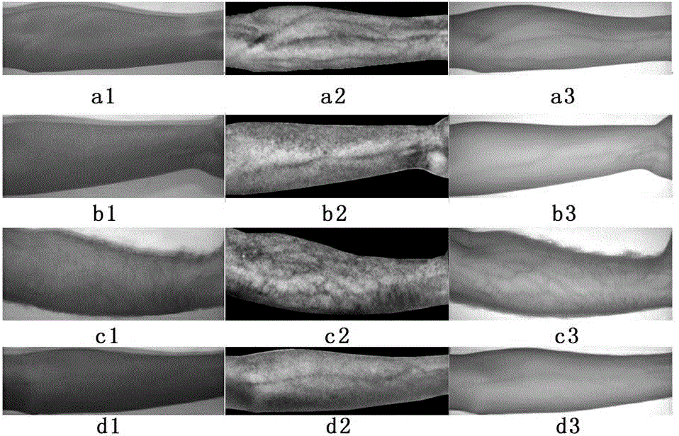
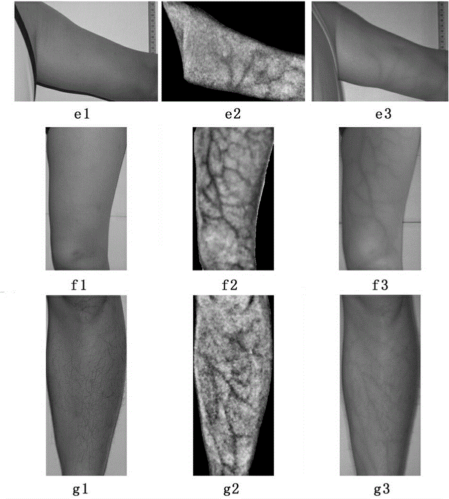
[0045] 1. Use the JAI-AD080CL visible light / near-infrared synchronous camera to capture images of the arm. The camera simultaneously captures the visible / near-infrared spectrum of arm images, with visible and near-infrared light passing through a single lens and entering two photosensitive chips. Among them, the first chip uses Bayer color technology, which only acquires visible light, that is, conventional images; the second chip is a monochromatic near-infrared imaging chip, and under the irradiation of a near-infrared light source, the near-infrared skin image taken by it can be See where the veins are; the two images are perfectly in sync. In order to improve the positioning accuracy of vein location, this example first collects images of the inner forearm of 20 people (each person has 1 group of visible light / near-infrared images, a total of 20 groups), and ...


PUM


Abstract

The invention discloses a fast vein imaging method for visible-light skin images, and belongs to the field of information perception and recognition. The method comprises steps as follows: N groups of skin images comprising visible-light images and near-infrared images which are completely synchronous are collected to build a skin image library; corresponding pixel blocks of each group of the visible-light images and near-infrared images are sequentially selected to form a training database; a three-layer feedforward neural network is adopted to simulate a mapping relation of visible-light pixels to near-infrared pixels in the training database, and training adjustment is performed; an RGB (red green blue) pixel value of a to-be-measured visible-light skin image is input to the three-layer feedforward neural network which is well trained so as to obtain vein imaging of the image. According to the pixel corresponding relation of visible-light and near-infrared synchronous images, the mapping between the visible-light and near-infrared synchronous images is realized by adopting the feedforward neural network, and then the vein imaging is realized through an energy result graph; the processing process is simple and easy to realize, and the method has high practical and popularizing value; extra special device and equipment are not required, and the imaging cost is greatly reduced.

Description

technical field [0001] The invention relates to a vein imaging method for skin images, in particular to a quick vein imaging method for visible skin images, which belongs to the field of information perception and recognition. Background technique [0002] More than a hundred years ago, human medicine advanced to the point of using venipuncture to draw blood and inject treatment. The key to implementing this medical technology is to first try to find the veins of the human body before needle puncture can be performed for follow-up treatment. At present, there are mainly two methods for locating veins in clinical use: visual method—finding and locating veins with naked vision. This method is only suitable for patients with superficial and prominent veins. The size, curvature and bifurcation of the veins cannot be clearly seen at a glance; touch method-find and locate veins with the feeling of touch. This method is based on many years of experience of medical staff, through t...

Claims


Application Information

Patent Timeline
IPC IPC(8): A61B5/00G06K9/00
CPCA61B5/0059A61B5/0077
Inventor 唐超颖王彪
Owner NANJING UNIV OF AERONAUTICS & ASTRONAUTICS
Learn More