
Multi-channel nerve repair conduit with tissue induced function and mold

A nerve repair and inductive technology, applied in the medical field, can solve problems such as easy inactivation, influence on nerve regeneration, and differences in biocompatibility, and achieve good degradability and biocompatibility, repair peripheral nerve defects, and mechanical strength Guaranteed effect

Active Publication Date: 2015-03-11
甘肃伯骊江3D打印科技有限公司
View PDF3 Cites 24 Cited by
  • Summary
  • Abstract
  • Description
  • Claims
  • Application Information

AI Technical Summary

Problems solved by technology

Whether the traditional first-generation tissue-engineered nerve conduits are natural materials or artificial materials, the main function is to guide nerve fiber regeneration. The main problems are: 1) Different materials have different biocompatibility; 2) The degradation performance of materials is different, which affects the regeneration of nerves; 3) it is easy to form scar structures, which seriously affects the biological functions of regenerated nerves; 4) the synthetic polymer materials lack cell recognition sites
However, the biggest problem facing this type of material is that the material lacks tissue induction (tissue induction refers to a new type of biomaterial that can directly induce the regeneration of living tissue. , and can induce cells to form tissue) functions, cannot fully simulate the tissue microenvironment suitable for cells and hosts, and is still far from ideal materials in terms of nerve regeneration and repair
[0005] The main problem faced by tissue-inducing nerve repair materials is that although materials constructed by compounding nerve-related growth factors, gene carriers or slow-release methods have tissue-inducing functions, the compounded active factors have unstable performance, easy inactivation and Shortcomings such as sudden release cannot achieve the goal of active repair function in the true sense

Method used

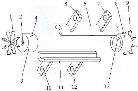
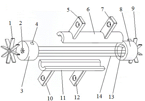
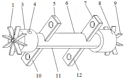

Image

  • Multi-channel nerve repair conduit with tissue induced function and mold
  • Multi-channel nerve repair conduit with tissue induced function and mold
  • Multi-channel nerve repair conduit with tissue induced function and mold

Examples


Embodiment 1

[0071] According to the mass ratio of 1:5, salidroside and chitosan were respectively taken, mixed uniformly, and chitosan-coated salidroside microspheres were prepared by using the tripolyphosphate method; the microspheres were treated with 1% genipin Cross-link at 37°C for 1 hour, freeze-dry in vacuum for later use; dissolve polylactic acid-polyglycolic acid copolymer in chloroform to obtain a saturated solution, add 10 mg of the prepared microspheres, stir with a magnetic stirrer at 50 rpm for 2 hours, and place in a fume hood Remove chloroform by volatilization, centrifuge at 4°C and 15,000rpm for 10 minutes, add appropriate amount of distilled water to disperse to obtain composite salidroside sustained-release microspheres, dry at room temperature for later use; use light microscope, laser particle size analyzer and scanning electron microscope to evaluate composite rhodiola respectively Morphology, particle size characteristics and characterization of glucoside sustained-...

Embodiment 2

[0073]According to the mass ratio of 1:10, salidroside and chitosan were respectively taken, mixed evenly, and chitosan-coated salidroside microspheres were prepared by using the tripolyphosphate method; the microspheres were treated with 1% genipin Cross-link at 37°C for 1 hour, vacuum freeze-dry for later use; dissolve polylactic acid-polyglycolic acid copolymer in chloroform to obtain a saturated solution, add 10 mg of the prepared microspheres, stir with a magnetic stirrer at 60 rpm for 2 hours, and place in a fume hood Remove chloroform in the medium, centrifuge at 4°C and 15000rpm for 10min, add appropriate amount of distilled water to disperse to obtain composite salidroside sustained-release microspheres, dry at room temperature for later use; use light microscope, laser particle size analyzer and scanning electron microscope to evaluate composite rhodiola respectively Morphology, particle size characteristics and characterization of glucoside sustained-release microsph...

Embodiment 3

[0075] According to the mass ratio of 1:50, salidroside and chitosan were respectively taken, mixed evenly, and chitosan-coated salidroside microspheres were prepared by using the tripolyphosphate method; the microspheres were treated with 1% genipin Cross-link at 37°C for 1 hour, vacuum freeze-dry for later use; dissolve polylactic acid-polyglycolic acid copolymer in chloroform to obtain a saturated solution, add 10 mg of microspheres, stir with a magnetic stirrer at 55 rpm for 2 hours, and volatilize in a fume hood Remove chloroform, centrifuge at 4°C and 15,000 rpm for 10 minutes at high speed, add appropriate amount of distilled water to disperse to obtain composite salidroside sustained-release microspheres, dry at room temperature for later use; use light microscope, laser particle size analyzer and scanning electron microscope to evaluate composite rhodiola Morphology, particle size characteristics and characterization of glucoside sustained-release microspheres; in vitr...


PUM


Abstract

The invention discloses a multi-channel nerve repair conduit with a tissue induced function and a mold; chitosan-coated salidroside microspheres and composite type salidroside slow-release microspheres are prepared, the drug cumulative release amount is calculated, the composite type salidroside slow-release microspheres with different contents are mixed with I-type collagen, and salidroside slow-release microspheres / I-type collagen is obtained; a multi-aperture cylindrical nerve conduit core layer is prepared by using the mold, a nano fiber nerve conduit shell layer is prepared by a high-pressure electrostatic spinning technology, the core layer and the shell layer are nested, and thus the multi-channel nerve repair conduit with the tissue induced function is prepared. The shell layer of the conduit has a good role in exchanging with a tissue fluid, and has a function of guiding nerve growth; the core layer has the functions of guiding nerve fiber orientation growth, promoting stem cells to directionally differentiate to schwann cells and accelerating the nerve fiber growth and function recovery, effectively repairs peripheral nerve defects, has good degradation and biocompatibility, and meets the requirements of a tissue engineering scaffold material.

Description

technical field [0001] The invention belongs to the technical field of medicine, and relates to a nerve repair catheter, in particular to a multi-channel nerve repair catheter with tissue inductive function; the invention also relates to a mold used in the preparation process of the multi-channel nerve repair catheter. Background technique [0002] Peripheral nerve injury is one of the most common clinical injuries. With the development of modern construction industry, transportation industry and the frequent occurrence of major natural disasters and local wars, the incidence of peripheral nerve injury is increasing year by year. Peripheral nerve injury often causes degeneration and dysfunction in the innervated area, which affects the physical and mental health and quality of life of patients for a long time, and has become one of the challenges facing the world's medicine. [0003] The use of tissue engineering methods to construct different nerve conduits through natural...

Claims


Application Information

Patent Timeline
IPC IPC(8): A61L29/04A61L29/06A61L29/16
Inventor 赵红斌甄平董菊子李晓云孙宏斌李根王九娜唐俊杰赵玲
Owner 甘肃伯骊江3D打印科技有限公司
Learn More