
Biological tissue opto-acoustic confocal micro-imaging device and method

A technology of confocal microscopy and imaging device, applied in the field of medical imaging, can solve the problems of low contrast and low resolution of photoacoustic microscopy imaging, and achieve the effect of increasing signal-to-noise ratio and image contrast

Inactive Publication Date: 2014-08-06
汇佳生物仪器(上海)有限公司
View PDF8 Cites 19 Cited by
  • Summary
  • Abstract
  • Description
  • Claims
  • Application Information

AI Technical Summary

Problems solved by technology

[0005] In order to solve the problem of low resolution and low contrast of photoacoustic microscopic imaging of biological tissues such as subcutaneous blood vessels, tumors, animal organs, etc. Detection sensitivity and image signal-to-noise ratio to obtain clear 3D images

Method used


Image

  • Biological tissue opto-acoustic confocal micro-imaging device and method
  • Biological tissue opto-acoustic confocal micro-imaging device and method

Examples


Embodiment Construction

[0021] Below in conjunction with accompanying drawing, the present invention will be further described:

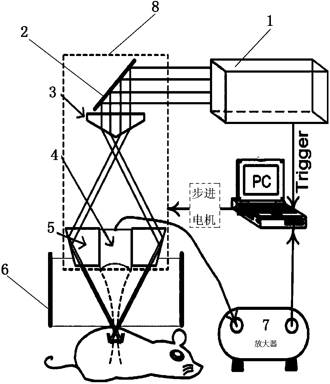
[0022] The present invention proposes a photoacoustic confocal microscopic imaging device for biological tissue, such as figure 1 As shown in , it is mainly composed of an imaging device, a signal acquisition device and a control device. It is characterized in that: both the imaging device and the signal acquisition device are connected to the control device, and the imaging device is connected to the signal acquisition device; Shaped lens 3, ultrasonic detector 4, optical condenser 5 and water tank 6; The outside of laser device 1 is provided with reflector 2 corresponding to the laser emission direction, below reflector 2 is provided with conical lens 3, and below conical lens 3 An ultrasonic detector 4 is arranged above the inside of the water tank 6 , and optical concentrators 5 are arranged on both sides of the ultrasonic detector 4 .

[0023] The control device is c...


PUM


Abstract

The invention relates to a biological tissue opto-acoustic confocal micro-imaging device and method, and belongs to the field of medical imaging. The biological tissue opto-acoustic confocal micro-imaging device is mainly composed of an imaging device, a signal collecting device and a control device. The imaging device and the signal collecting device are connected with the control device, and the imaging device is connected with the signal collecting device. The biological tissue opto-acoustic confocal micro-imaging device is simple and reasonable in structure, a clear three-dimensional image is obtained by improving the detection sensitivity and the image signal to noise ratio, the problems that an opto-acoustic micro-image of the biological tissue such as the subcutaneous blood vessels, tumors and animal organs is low in resolution ratio and contrast ratio under the out-of-focus situation are solved, and the biological tissue opto-acoustic confocal micro-imaging device and method are suitable for popularization and application.

Description

technical field [0001] The invention belongs to the field of medical imaging, and in particular relates to a photoacoustic confocal microscopic imaging device and method capable of performing three-dimensional clear imaging of subcutaneous blood vessels, tumors, animal organs and the like. Background technique [0002] Photoacoustic imaging technology is a new type of medical imaging technology that has developed rapidly in recent years. It adopts the method of "photo-induced acoustic detection image reconstruction" for imaging. It uses the endogenous optical absorption properties of the sample for imaging, so it can obtain structural and functional images of tissue samples, and can also obtain the three-dimensional distribution of optical energy deposition. As a non-invasive imaging technology, photoacoustic imaging not only has the characteristics of high resolution and high contrast of optical imaging technology, but also has the characteristics of high penetration depth ...

Claims


Application Information

Patent Timeline
Patent Type & Authority Applications(China)
IPC IPC(8): A61B5/00A61B8/00
Inventor 聂立铭陈小元
Owner 汇佳生物仪器(上海)有限公司
Learn More