
Fish skin collagen support loading epidermal growth factors and preparation method thereof

A technology of epidermal growth factor and collagen scaffold, applied in the field of medical devices, can solve the problems of rapid diffusion of epidermal growth factor, virus contamination, loss of biological activity, etc. short effect

Inactive Publication Date: 2015-07-22
FUZHOU UNIV
View PDF4 Cites 0 Cited by
  • Summary
  • Abstract
  • Description
  • Claims
  • Application Information

AI Technical Summary

Problems solved by technology

[0004] At present, the main way to treat diabetic ulcers is to directly supplement exogenous epidermal growth factor locally on the wound surface, but this method of administration has the following disadvantages: the epidermal growth factor diffuses quickly and has a short half-life. Without carrier protection, it is easy to be diluted or Degradation and loss of biological activity, manifested as the topical epidermal growth factor water agent can be degraded and destroyed by the protease in the wound exudate within a few minutes after contacting the skin wound, or it can be quickly lost from the wound
At present, the collagen used in industry and clinical application is extracted from terrestrial animals such as cattle, sheep, pigs, etc., and this type of collagen has the risk of being contaminated by viruses such as mad cow disease and foot-and-mouth disease carried by animals.

Method used


Image

  • Fish skin collagen support loading epidermal growth factors and preparation method thereof
  • Fish skin collagen support loading epidermal growth factors and preparation method thereof

Examples


Embodiment 1

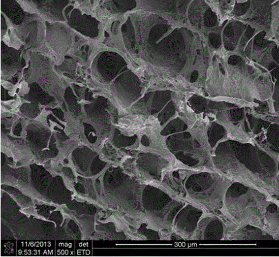
[0022] Embodiment 1: the preparation of fish skin collagen scaffold

[0023] (1) Thaw the fish skin and cut it into pieces;

[0024] (2) After washing with distilled water, add a mixed solution of hydrogen peroxide with a volume fraction of 1% and 0.01 mol / L sodium hydroxide that is 20 times its mass, stir mechanically for 24 hours, and replace the alkaline solution every 8 hours;

[0025] (3) Add isopropanol solution with a volume fraction of 10%, mechanically stir for 4 h, and wash with purified water;

[0026] (4) Add 2.5% sodium chloride solution, stir mechanically for 12 h, and wash with purified water;

[0027] (5) Add 10 volumes of purified water, adjust pH=2.0-3.0 with hydrochloric acid, add pepsin with 2.5% fish skin mass, enzymolyze at 4-8°C for 16-24 h, centrifuge at 8000-20000 rpm for 20 min, take Serum;

[0028] (6) Add a certain amount of sodium chloride to the above supernatant to a final salt concentration of 0.9mol / L, salt out, centrifuge at 8000 rpm for 20...

Embodiment 2

[0031] Example 2: Preparation of Nanoparticles Loaded with Epidermal Growth Factor

[0032] (1) Dissolve chitosan in 1% acetic acid, dialyze for 4 days, and freeze-dry to obtain purified chitosan;

[0033] (2) Dissolve 0.2 g chitosan in 5 ml 1% acetic acid, adjust the pH value to 6.0 with 0.1mol / L NaOH, and set the volume to 100 ml to obtain a chitosan solution with a concentration of 2 mg / ml;

[0034] (3) Prepare a heparin solution with a concentration of 1 mg / ml, and use this solution as the mother solution to prepare a heparin mixture solution containing epidermal growth factor 10500 IU / ml;

[0035] (4) At 4°C, under electric stirring, drop 2ml of the 1mg / mL above heparin mixture into 5ml of the 2 mg / ml chitosan solution drop by drop at a rate of 10-40 drops per minute to obtain dispersion Uniformly loaded nanoparticles of epidermal growth factor.

Embodiment 3

[0036] Example 3: Preparation of fish skin collagen scaffold loaded with epidermal growth factor

[0037] The fish skin collagen scaffold prepared in Example 1 was infiltrated with an aqueous solution of nanoparticles loaded with epidermal growth factor at a ratio of 2 ml / cm 2; Store at -20°C after infiltration. Vacuum freeze-drying, that is.


PUM

PropertyMeasurementUnit
quality scoreaaaaaaaaaa

Abstract

The invention discloses a fish skin collagen support loading epidermal growth factors and a preparation method thereof. The fish skin collagen support is composed of cross-linked fish skin collagen support bodies and nanometer particles of the load epidermal growth factors. The preparation method comprises the steps that after dewatering, degreasing, enzyme treatment and dialysis is conducted on fish skin, the cross-linked fish skin collagen support bodies are prepared, then the cross-linked fish skin collagen support bodies are soaked in a solution of the nanometer particles of the load epidermal growth factors, and the fish skin collagen support loading the epidermal growth factors is obtained after freezing and drying. The support has a slow releasing effect on the loaded factors, and prolongs the acting time of the epidermal growth factors at a trauma position. The fish skin collagen support obtained through the method has the advantages of being good in biocompatibility and biodegradability, free of immunogenicity and the like, is harmless to a wound surface, can effectively induce wound surface healing and is a good medical material.

Description

technical field [0001] The invention belongs to the technical field of medical devices, and in particular relates to a fish skin collagen scaffold loaded with epidermal growth factor for treating diabetic ulcers and a preparation method thereof. Background technique [0002] With the improvement of living standards, changes in life patterns and the aging of society, the incidence of diabetes is increasing year by year. Diabetic ulcer is one of its common complications. It is due to vascular and neuropathy caused by diabetes, which causes abnormal changes in the skin. The clinical manifestations are local erosion, ulcer, necrosis, and the wound surface does not heal for a long time. , untimely treatment will cause disease spread, surgical amputation (toe), and even life-threatening. Diabetic ulcers often recur and persist, forming stubborn and refractory ulcers, which seriously endanger the health of diabetic patients, reduce the quality of life, and bring great social and e...

Claims


Application Information

Patent Timeline
Patent Type & Authority Patents(China)
IPC IPC(8): A61L27/24A61L27/54A61L27/60A61L27/20
Inventor 张其清贺超龙王建华程娘梅杨秋陈名懋伍久林
Owner FUZHOU UNIV
Learn More