
Methods for separating and identifying DNA (deoxyribonucleic acid) binding protein through DNA co-immunoprecipitation

A technology for binding proteins and DNA molecules, which is applied in the field of separation and identification of DNA binding proteins to achieve the effect of improving experimental efficiency, stability and repeatability

Active Publication Date: 2014-04-23
CANCER INST & HOSPITAL CHINESE ACADEMY OF MEDICAL SCI +1
View PDF1 Cites 8 Cited by
  • Summary
  • Abstract
  • Description
  • Claims
  • Application Information

AI Technical Summary

Problems solved by technology

However, there is still a lack of methods to use known DNA sequences to isolate proteins that specifically bind to the nucleus and further identify DNA-binding proteins.

Method used


Image

  • Methods for separating and identifying DNA (deoxyribonucleic acid) binding protein through DNA co-immunoprecipitation
  • Methods for separating and identifying DNA (deoxyribonucleic acid) binding protein through DNA co-immunoprecipitation
  • Methods for separating and identifying DNA (deoxyribonucleic acid) binding protein through DNA co-immunoprecipitation

Examples


Embodiment 1

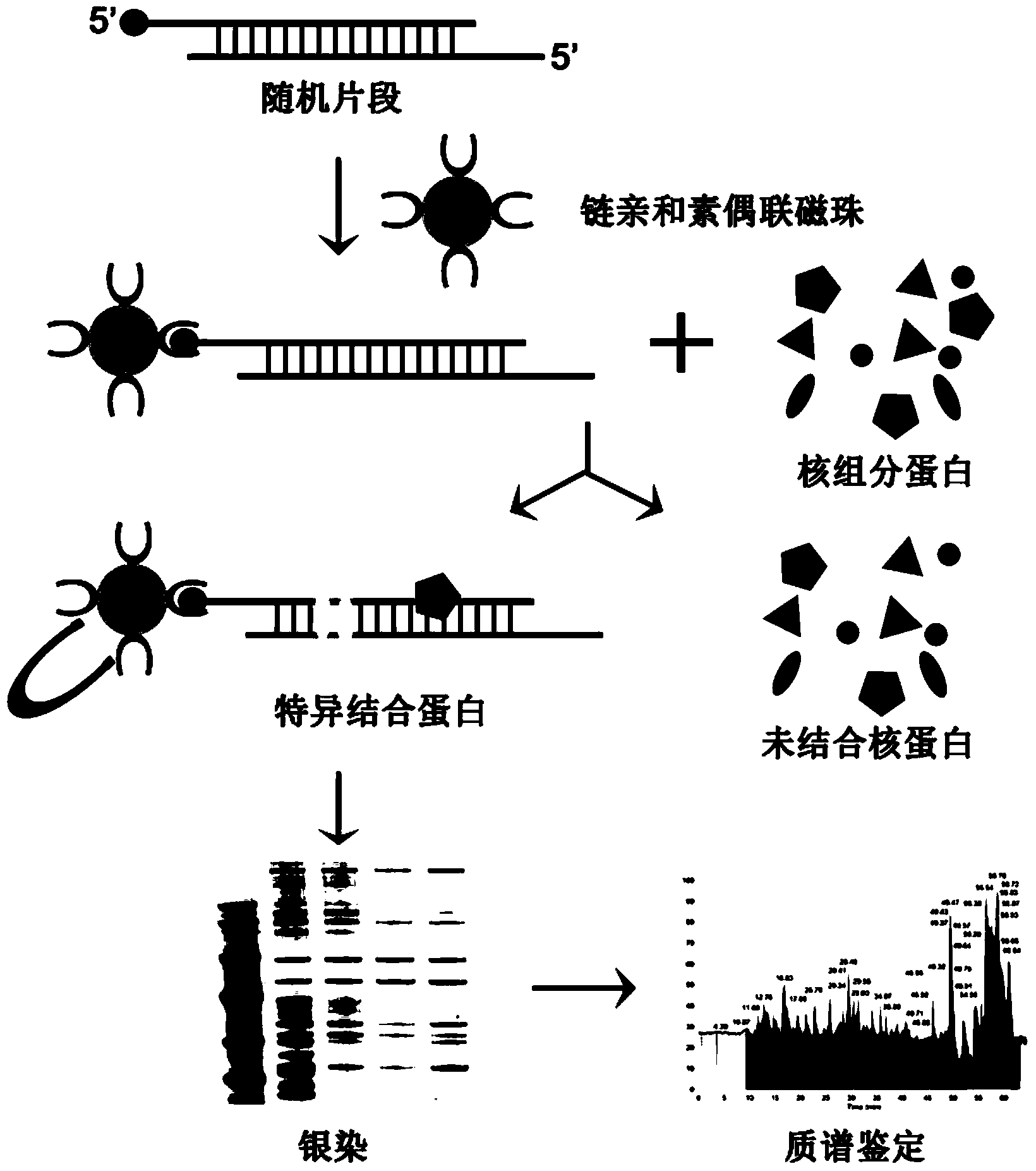
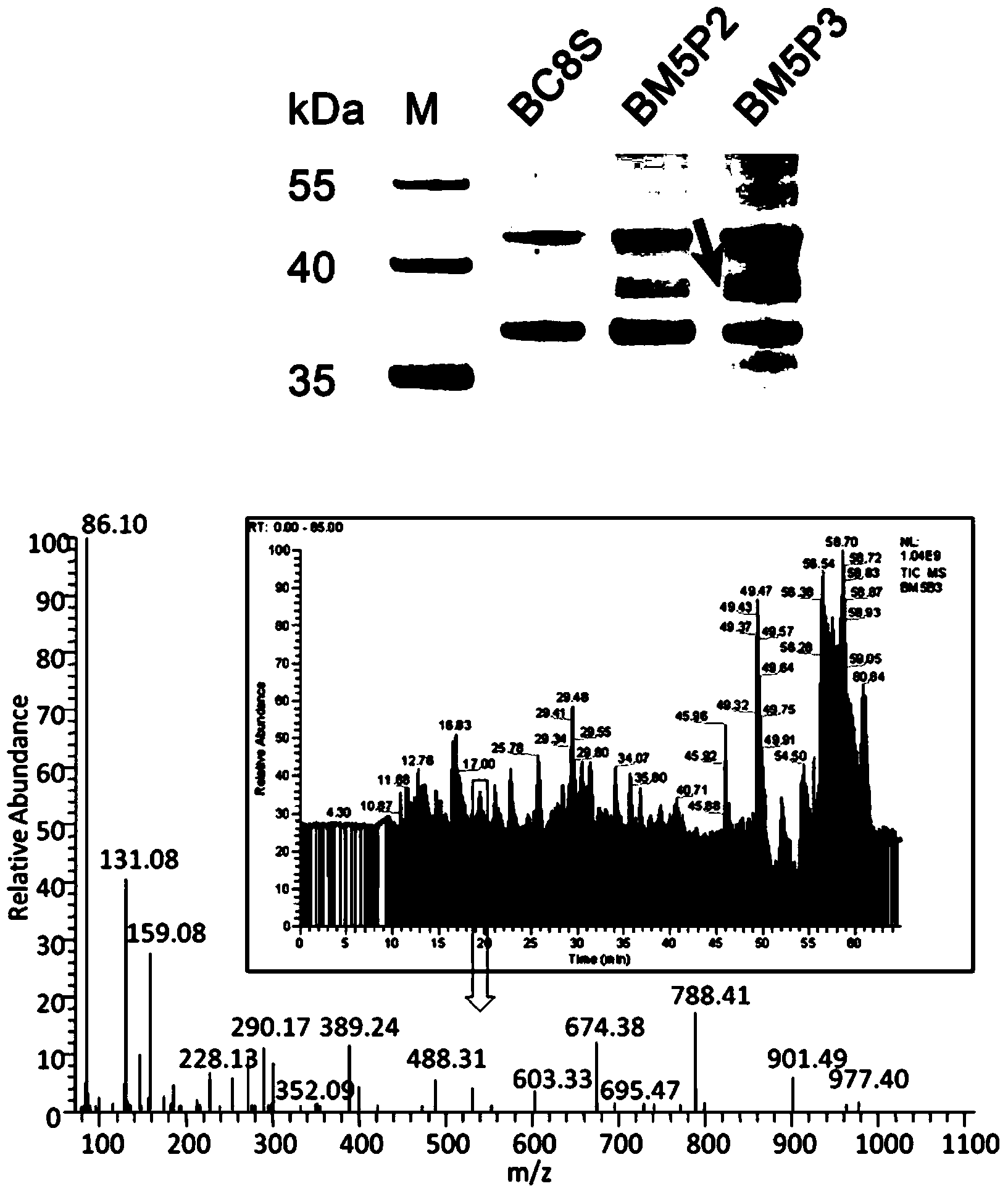
[0055] Example 1: Identification of PURA transcription factors using DNA co-immunoprecipitation combined with mass spectrometry

[0056] 1. Extraction of Nuclear Component Proteins

[0057] 1) Culture esophageal cancer cells KYSE510 (gifted by Professor Shimada of Kyoto University, Japan) in a T75 culture flask, and wash the cells with 4°C pre-cooled 1×PBS, 10ml / time×1 time. After digestion with trypsin, stop the translation of trypsin with 10ml of complete medium (RPMI1640, containing 10% fetal bovine serum), wash the cells with 10ml of PBS / time×2 times, discard the PBS and leave the precipitated cells; use 1mL Buffer A (10mM HEPES ,pH7.9,1.5mM MgCl 2 , 10mM NaCl, 0.5mM DTT) to resuspend the cells and place on ice for 15min;

[0058] 2) Add a final concentration of 0.3% NP40 lysate (50mM Tris (pH7.4), 150mM NaCl, 1% NP-40) to the cell suspension in step 1), mix well and add the cell suspension to the homogenizer , homogenate up and down 20 times;

[0059] 3) After homogen...

Embodiment 2

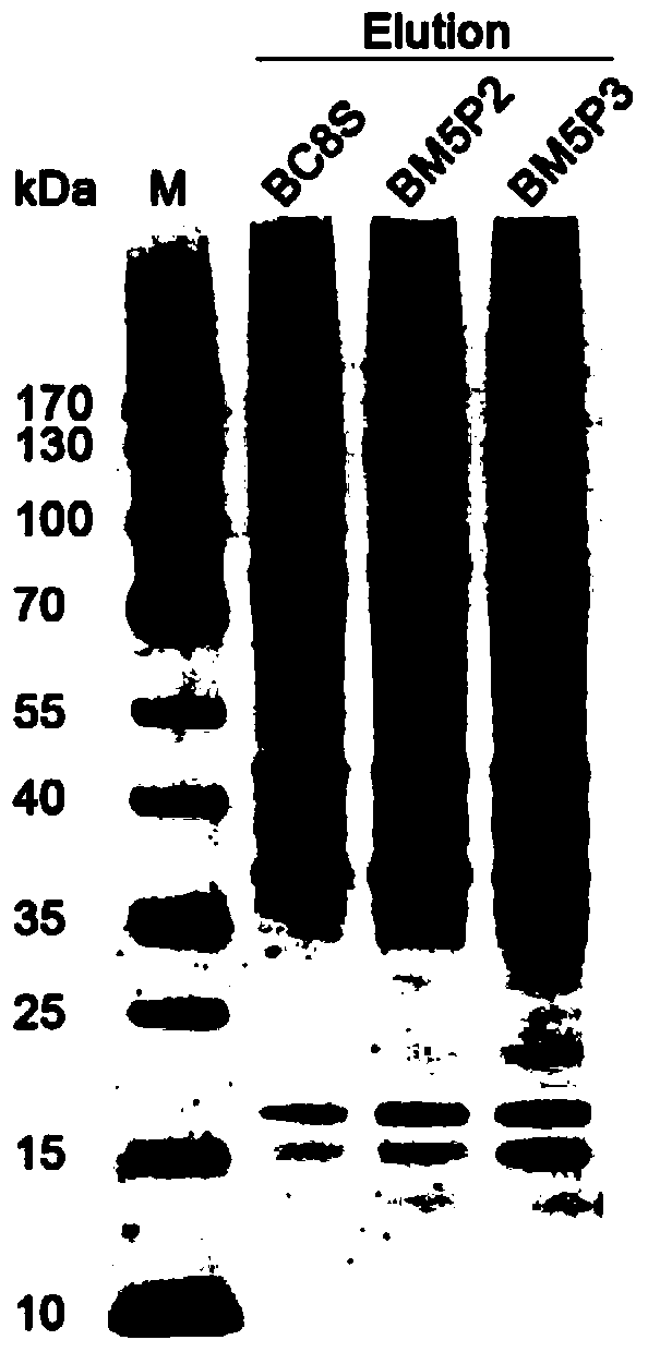
[0107] Example 2: Using DNA co-immunoprecipitation technology Western blotting to identify PARP transcription factors

[0108] 1. Extraction of Nuclear Component Proteins

[0109] Use a T75 culture flask to culture KYSE510 cells, wash the cells with 4°C pre-cooled 1×PBS, 10ml / time×1 time. After digestion with trypsin, stop the translation of trypsin with 10ml of complete medium, wash the cells with 10ml / time×2 times of PBS, discard the PBS and leave the precipitated cells; use 1mL BufferA (10mM HEPES, pH7.9, 1.5mM , 0.5mM DTT) to resuspend the cells and place on ice for 15min;

[0110] 1) Add NP40 lysate to the cell suspension in step 1) to a final concentration of 0.3%, mix well, add the cell suspension into a homogenizer, and homogenize up and down 20 times;

[0111] 2) After homogenization, the cell lysate suspension was reduced to 228g, centrifuged at 4°C for 10min, and the supernatant was discarded;

[0112] 3) Resuspend the nucleus pellet with 300μl Buffer B (0.3M suc...


PUM


Abstract

The invention relates to methods for separating and identifying DNA (deoxyribonucleic acid) binding protein, in particular to methods for separating and identifying DNA binding protein through a DNA co-immunoprecipitation technology. The separating method comprises the steps of extracting nucleoprotein of a cell, mixing a target DNA molecule and nucleoprotein, adding a molecular entity capable of being bound with the target DNA molecule, separating to obtain a nucleoprotein ingredient bound with the target DNA molecule, further separating the obtained nucleoprotein ingredient, and obtaining the DNA binding protein. The identifying method further comprises the step of identifying the DNA binding protein obtained by separating. The methods have the characteristics of good stability and repeatability, and significantly improve the experiment efficiency of identifying the unknown DNA binding protein. The methods provide beneficial help for subjects such as molecular behavioristics, molecular oncology, cytobiology and biochemistry deeply researching into cytogene transcriptional control and genetic transcription.

Description

technical field [0001] The invention relates to a method for separating and identifying DNA binding proteins, in particular to a method for separating and identifying DNA binding proteins using DNA co-immunoprecipitation technology. Background technique [0002] Biomedical research has evolved from the hypothesis of one gene and one protein to analyzing the functions of many genes or proteins at the omics level. Due to the complexity of various life activities of cells, the sequence information of the whole genome cannot explain the biological functions of cells, and the final product of genes is the ultimate executor of cell composition and function. In the process of cell life, the opening and closing of specific genes is a complex system engineering, and the transcriptional regulation of human genes has always been a hot research field for biologists. At present, chromatin immunoprecipitation (ChIP) is mainly used to study the interaction between known protein transcript...

Claims


Application Information

Patent Timeline
IPC IPC(8): C07K1/14C07K1/26G01N27/62
Inventor 赵晓航许杨林正伟郭志敏赵楠
Owner CANCER INST & HOSPITAL CHINESE ACADEMY OF MEDICAL SCI
Learn More