Method for simultaneously segmenting liver and blood vessel in CTA (computed tomography angiography) image

An image, liver technology, applied in the field of medical image processing, can solve problems such as difficulty in segmenting the liver

Active Publication Date: 2014-04-02
ZHEJIANG DE IMAGE SOLUTIONS CO LTD
View PDF3 Cites 8 Cited by
  • Summary
  • Abstract
  • Description
  • Claims
  • Application Information

AI Technical Summary

Problems solved by technology

In addition, since the abdominal scan image contains many irrelevant tissues, such as stomach, heart, spleen, etc., which are very close to the gray value of the liver, it also contains blood vessels, which makes it difficult to directly extract blood vessels and segment the liver in complex images. very difficult

Method used


Image

  • Method for simultaneously segmenting liver and blood vessel in CTA (computed tomography angiography) image
  • Method for simultaneously segmenting liver and blood vessel in CTA (computed tomography angiography) image
  • Method for simultaneously segmenting liver and blood vessel in CTA (computed tomography angiography) image

Examples


Embodiment Construction

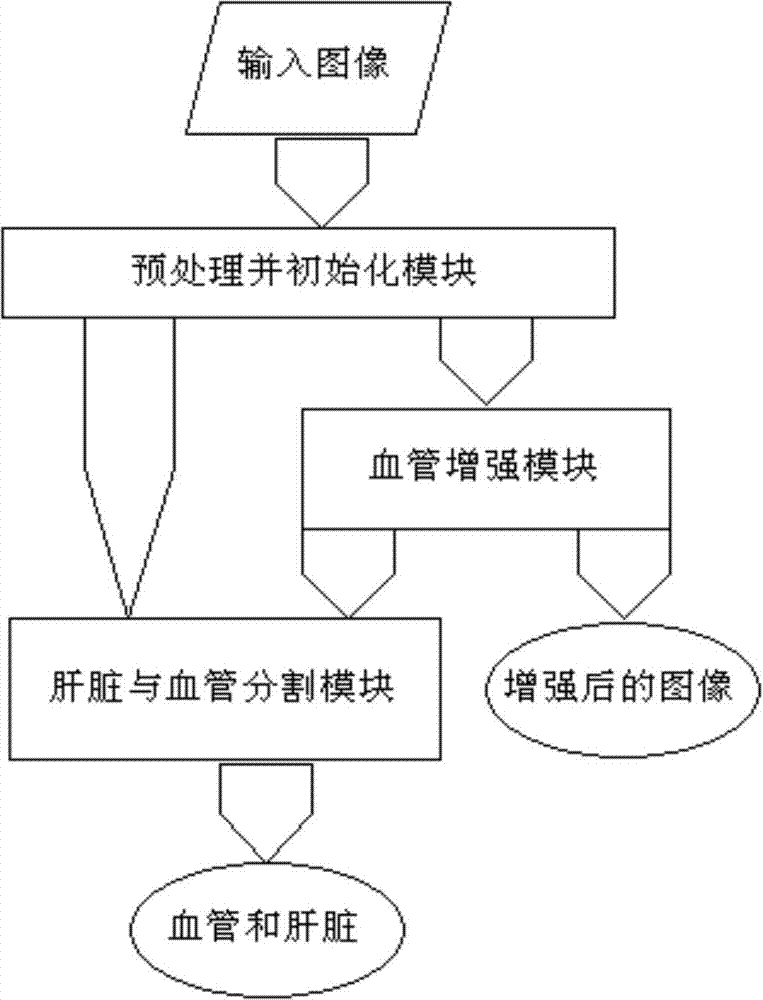
[0067] figure 1 The process of blood vessel enhancement and liver and blood vessel segmentation in the CTA scan image is shown in the figure. The specific process is as follows:

[0068] In the implementation process, the input of blood vessel and liver segmentation can be an image enhanced with blood vessels, or an image without enhancement, and the former is adopted in this embodiment.

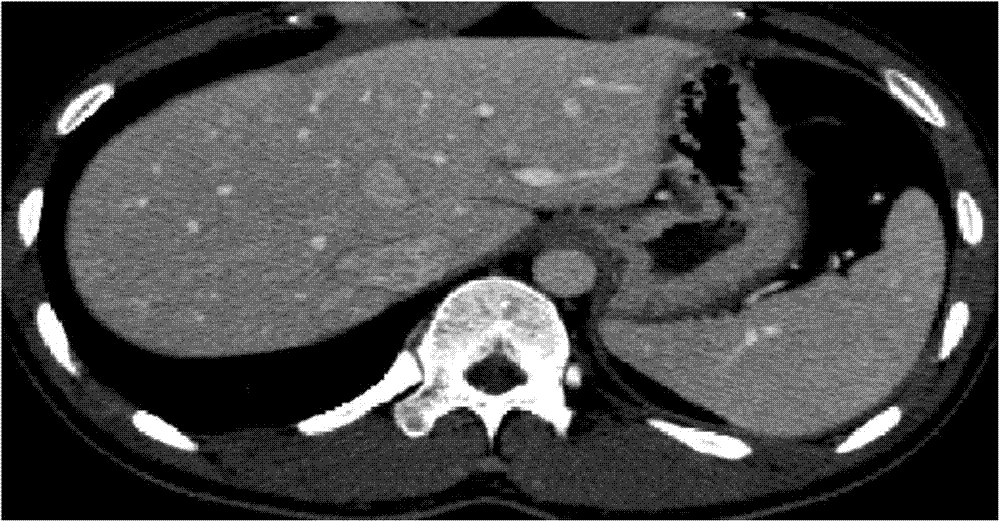
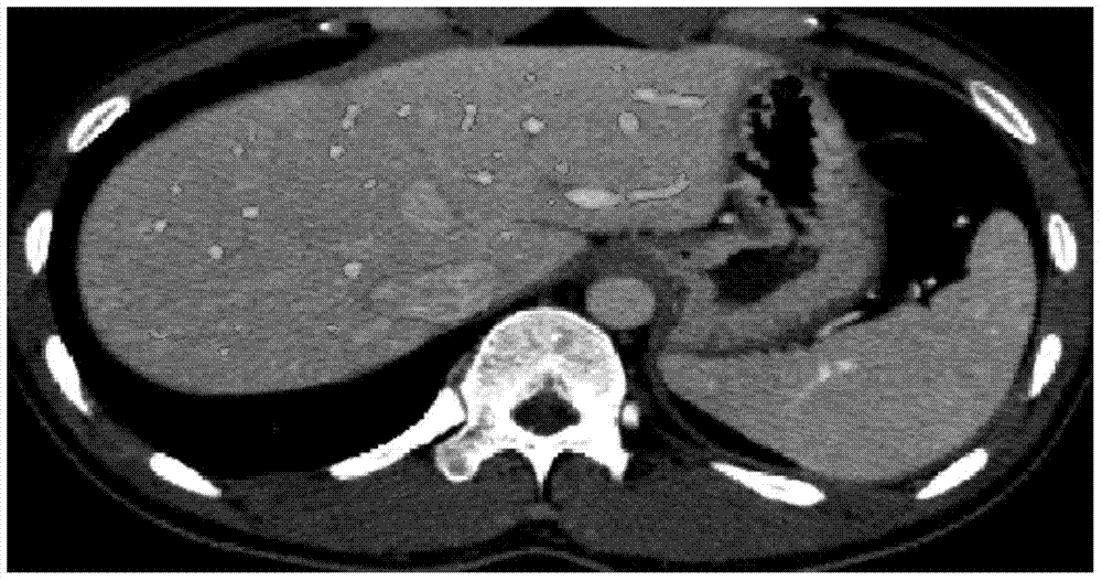
[0069] 1. Input liver CTA or MRA scan image I 1 , the size is 512×512×368, and the window width and level are adjusted so that the gray scale range of the liver and blood vessels is mainly between 0 and 255. figure 2 It is the 88th slice image of the three-dimensional liver data cross section. Perform Gaussian denoising on the image: I=I 1 *G δ , * is the convolution operator, is a Gaussian kernel function with window δ. In this example, δ=0.5. The initialization adopts interactive software, and randomly selects a liver area without blood vessels inside the liver.

[0070] 2. In an...


PUM


Abstract

The invention relates to medical image processing, and aims to provide a method for simultaneously segmenting liver and a blood vessel in a CTA (computed tomography angiography) image. The method comprises the following steps of (1) performing model initialization; (2) optimizing a new variational energy model, and simultaneously segmenting the liver and the blood vessel; (3) performing post-processing to obtain a liver contour and a blood vessel contour respectively. According to the method for simultaneously segmenting the liver and the blood vessel, the adverse impact of weak boundaries, noise and the like can be effectively overcome; particularly, due to the introduction of gray level distribution information of local neighborhoods, the liver can be well distinguished from adjacent organs or soft tissues, and the phenomenon of over-segmentation can be effectively prevented.

Description

[0001] This case application is a divisional application of the Chinese invention patent application "Method for Enhancement of Liver Vessels in CTA Images and Simultaneous Segmentation of Liver and Vessels". The original application date is January 17, 2012; the application number is 201210014249.9. technical field [0002] The invention relates to the field of medical image processing, in particular to the enhancement of liver blood vessels in CT (computed tomography contrast enhancement) images and the simultaneous segmentation of liver entities and blood vessels. Background technique [0003] The structure of the liver and blood vessels is of great significance for the diagnosis of liver disease and the guidance of liver disease surgery. CTA (computed tomography contrast-enhanced) technology is one of the most commonly used liver and blood vessel imaging diagnostic techniques. Through CTA, doctors can obtain a series of enhanced two-dimensional CT slices of blood vessels...

Claims


Application Information

Patent Timeline
Patent Type & Authority Applications(China)
IPC IPC(8): G06T5/00A61B6/03
Inventor 孔德兴彭佳林吴法王金伟
Owner ZHEJIANG DE IMAGE SOLUTIONS CO LTD
Why Eureka
Try Eureka