
Method for preparing electrospun artificial blood vessel with micro-nano biomimic intima structure

A technology of artificial blood vessels and electrospinning, applied in the direction of human tubular structure devices, blood vessels, etc., to achieve the effect of improving blood compatibility

Inactive Publication Date: 2013-07-24
王深明 +4
View PDF6 Cites 27 Cited by
  • Summary
  • Abstract
  • Description
  • Claims
  • Application Information

AI Technical Summary

Problems solved by technology

[0006] In view of the problems existing in the existing artificial blood vessel preparation technology, the main purpose of the present invention is to provide a method for preparing an electrospun artificial blood vessel with a micro-nano bionic intima structure, so as to improve the blood compatibility of the artificial blood vessel and avoid stenosis of artificial vessel lumen

Method used


Image

  • Method for preparing electrospun artificial blood vessel with micro-nano biomimic intima structure
  • Method for preparing electrospun artificial blood vessel with micro-nano biomimic intima structure
  • Method for preparing electrospun artificial blood vessel with micro-nano biomimic intima structure

Examples


Embodiment 1

[0039] Preparation of Polyurethane Artificial Blood Vessel with Micro-Nano Biomimetic Intima Structure

[0040] 1. Preparation of polyurethane solution

[0041] Dissolve polyurethane particles into a mixed solution of tetrahydrofuran and N,N-dimethylformamide with a volume ratio of 7:3, and stir for about 3 hours to form a clear, stable and uniform solution.

[0042] 2. Preparation of oriented electrospun fiber membrane

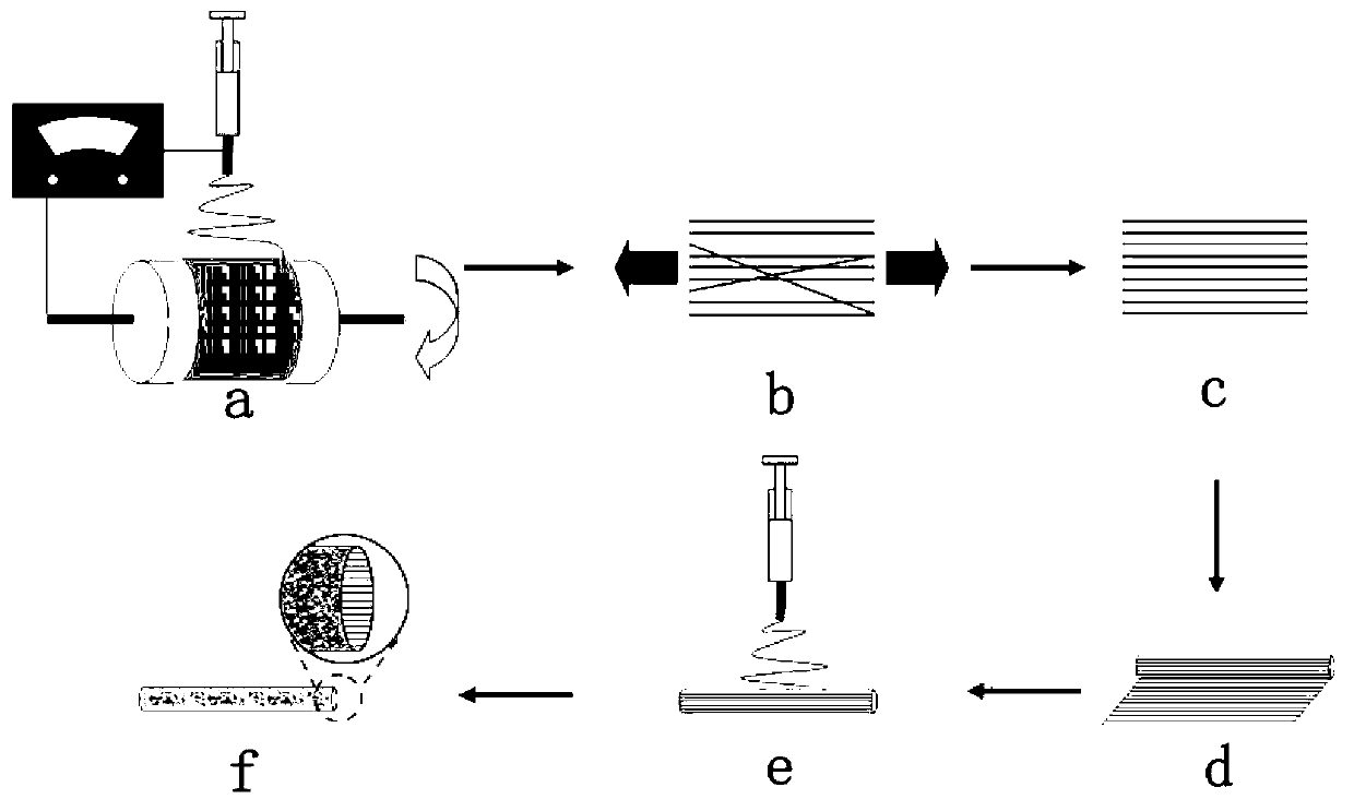
[0043] Move the solution into a 5ml syringe, then fix the syringe on the vertical support, connect the needle to the high voltage power supply, the distance between the needle and the upper end of the drum is 25cm, start the drum, set the speed to 3000rpm, and then apply 10-30KV After 30 minutes of high pressure, a film with an oriented fiber structure can be obtained.

[0044] 3. Stretching of oriented electrospun fiber membranes

[0045] In order to obtain a better degree of orientation, the oriented fiber film was stretched 120% along the long axis of th...

Embodiment 2

[0055] Preparation of polycaprolactone artificial blood vessel with micro-nano biomimetic intima structure

[0056] 1. Preparation of polycaprolactone solution

[0057] Dissolve polycaprolactone particles into a mixed solution of dichloromethane and N,N-dimethylformamide with a volume ratio of 5:5, and stir for about 3 hours to form a clear, stable and uniform solution.

[0058] 2. Preparation of oriented electrospun fiber membrane

[0059] Move the solution into a 5ml syringe, then fix the syringe on the vertical support, connect the needle to the high voltage power supply, the distance between the needle and the upper end of the drum is 27cm, start the drum, set the speed to 4000rpm, and then apply 10-30KV After 30 minutes of high pressure, a film with an oriented fiber structure can be obtained.

[0060] 3. Stretching of oriented electrospun fiber membranes

[0061] In order to obtain a better degree of orientation, the oriented fiber film was stretched 150% along the l...


PUM


Abstract

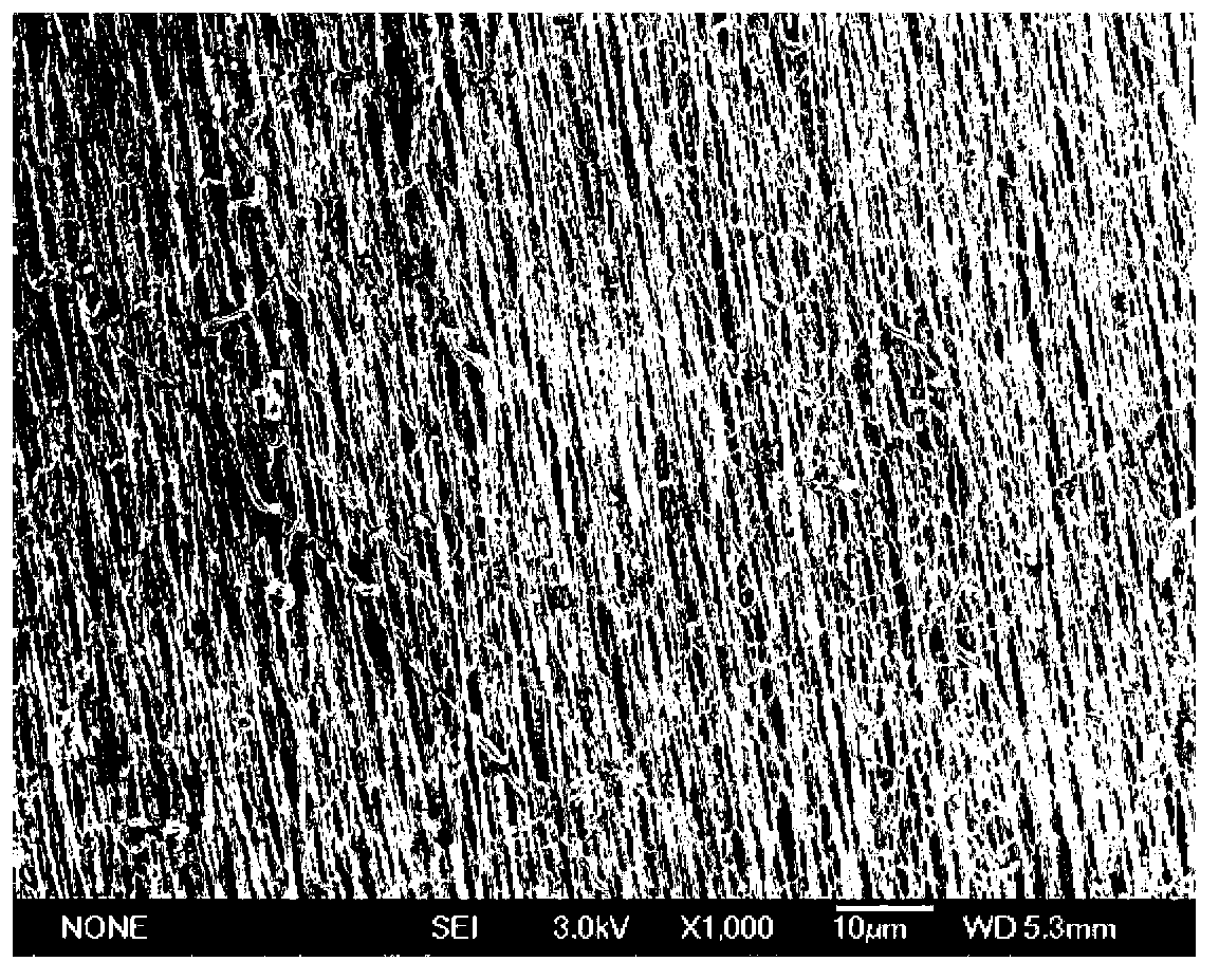
The invention discloses a method for preparing an artificial blood vessel with a micro-nano biomimic intima structure by utilizing an electrospinning technology. The method mainly comprises the following steps of preparing a macromolecular polymer solution; preparing an alignment electrospun fibrous membrane; stretching the alignment electrospun fibrous membrane; preparing an artificial blood vessel; performing characterization by using a scanning electron microscope; and determining blood compatibility. The artificial blood vessel prepared by the method has an intima imitation alignment microstructure, so that the blood compatibility of the artificial blood vessel meets a requirement on clinical anticoagulation performance, and a new way for the preparation of a novel artificial blood vessel is provided. In addition, the adoption of an anticoagulant is avoided, and the blood compatibility of the artificial blood vessel is improved only by a surface microstructure effect, so that side effects caused by the anticoagulant are reduced, and the method has broad application prospect in the field of medical clinical blood vessel transplantation.

Description

technical field [0001] The invention relates to a method for preparing an electrospun artificial blood vessel with a micro-nano bionic intima structure. Background technique [0002] At present, cardiovascular disease, cerebrovascular disease and peripheral arterial disease are still the main causes of human death worldwide. When diseased blood vessels need to be replaced, autologous blood vessels are the most commonly used clinically, such as saphenous vein, internal mammary artery, etc. About 600,000 vascular grafts need to be performed every year, but nearly one-third of the patients are due to aging blood vessels. Autologous blood vessels cannot be used due to lesions, injuries, or previous surgical use, and this method often requires a second operation, causing greater damage to the patient's body. In this case, artificial blood vessels become a good choice. Currently clinically used artificial blood vessels such as ePTFE ( ) and Dacron ( ) to replace large-diamet...

Claims


Application Information

Patent Timeline
Patent Type & Authority Applications(China)
IPC IPC(8): A61F2/06
Inventor 王深明刘瑞明赵勇胡作军秦原森
Owner 王深明
Learn More