
Adhesive cell scanning electron microscope carrier and preparation method

A technology of scanning electron microscopy and adherent cells, which is applied in the direction of circuits, discharge tubes, electrical components, etc., can solve the problems of large bonding force, time-consuming and laborious, shrinkage of adherent cells, etc., achieve good dehydration effect, maintain properties, and simple operation Effect

Active Publication Date: 2016-01-20
FIRST AFFILIATED HOSPITAL OF DALIAN MEDICAL UNIV
View PDF4 Cites 0 Cited by
  • Summary
  • Abstract
  • Description
  • Claims
  • Application Information

AI Technical Summary

Problems solved by technology

[0003] 1. Due to the poor cell attachment of glass slides, in order to improve cell attachment, the slides must be coated before culture, which is time-consuming and laborious;
[0004] 2. After multiple dehydration and drying treatments, glass slides become more brittle and easy to break, resulting in changes in cell morphology, especially the breakage of dendrites with longer dendrites; at the same time, the cell surface will be covered by lipid precipitation A thick film makes the outline of the adherent cells faintly visible, and in severe cases causes the adherent cells to shrink and collapse. All phenomena will affect the experimental results;
[0005] 3. The glass slide covered with the cell layer needs to be taken out of the cell culture dish every time. Because of the strong adhesion between the glass slide and the bottom of the culture dish, it is difficult to take out the slide and scratches are likely to occur on the cell surface;
[0006] 4. Difficulty distinguishing the cell side from the underside of the glass slide, sometimes resulting in mounting the cell side down glass slide on the sample holder
However, so far there is no relevant report on the direct preparation of SEM carriers for adherent cells using TC surface cell culture dishes

Method used


Image

  • Adhesive cell scanning electron microscope carrier and preparation method

Examples


Embodiment Construction

[0026] Follow the steps in turn:

[0027] a. Culture the cells to the desired density in a 35mmTC surface cell culture dish;

[0028] b. Fix the cells with glutaraldehyde with a pH of 7.2 to 7.4 and a concentration of 2.5%;

[0029] c. Wash the cells 3 times with 0.1mol / L PBS buffer, then soak the cells in 0.1mol / L PBS buffer for 12-15 hours;

[0030] d. Wash the cells once with 1×PBS buffer;

[0031] e. Add tert-butanol to the cells from low to high according to the concentration gradient. Every time tert-butanol is added, gently pipette 3 to 5 times, let it stand for 10 minutes, and wash the cells once with 1×PBS buffer until the last addition. Tert-butanol; after the last addition of tert-butanol, there is no need to blow and stand still, but to directly crystallize tert-butanol under a liquid nitrogen environment; the concentration gradient of said tert-butanol is 10%, 25%, 50% , 60%, 70%, 80%, 90%, 100%;

[0032] f. Put the TC surface cell culture dish into a vacuum m...


PUM


Abstract

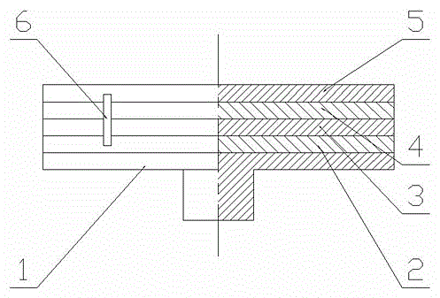
The invention discloses an adherent cell scanning electron microscope carrier which comprises a scanning electron microscope sample support (1). A base plate (3) of a tissue culture (TC) surface cell culture dish is fixed on the scanning electron microscope sample support (1) through an electric conduction adhesive layer (2), a cell layer (4) is coated on the base plate (3) of the TC surface cell culture dish, a conductive film (5) is arranged on the cell layer (4), and a conductive strip (6) is connected with the conductive film (5) and the electric conductive adhesive layer (2) in a crossed mode. According to the preparation method of the adherent cell scanning electron microscope carrier directly cultures cells by means of the TC surface cell culture dish, ethanol is replaced with tert butyl alcohol to be used as dehydrating agent, the adherent cell scanning electron microscope carrier is prepared by means of the base plate of the TC surface cell culture dish which is coated with the cell layer, and therefore, the adherent cell scanning electron microscope carrier and the preparation method have the advantages of being simple, rapid, convenient to use and the like.

Description

technical field [0001] The invention relates to an adherent cell scanning electron microscope carrier and a preparation method thereof, in particular to an adherent cell scanning electron microscope carrier and a preparation method which are simple in operation, low in cost, avoid cell shape changes, and ensure experimental effects. Background technique [0002] At present, in the medical field, it is very common to observe the surface morphology of adherent cells with a scanning electron microscope (electron microscope). Since the scanning electron microscope uses various physical signals excited by the finely focused electron beam to scan the sample surface to modulate imaging, the adherent cells must be prepared as a carrier for scanning electron microscopy, that is, the adherent cell scanning electron microscope carrier. Most of the existing adherent cell scanning electron microscope carriers use glass slides as culture plates. The preparation method is basically to put ...

Claims


Application Information

Patent Timeline
Patent Type & Authority Patents(China)
IPC IPC(8): H01J37/20
Inventor 宋智琦李乃浩
Owner FIRST AFFILIATED HOSPITAL OF DALIAN MEDICAL UNIV
Learn More