
Method for directional induction of differentiation of mesenchymal stem cells (MSCs)

A stem cell differentiation and mesenchymal stem cell technology, applied in the field of directional induction of mesenchymal stem cell differentiation, can solve problems such as cell separation difficulties, identification or application obstacles, and achieve the effect of improving induction efficiency

Inactive Publication Date: 2013-06-12
FIRST HOSPITAL AFFILIATED TO GENERAL HOSPITAL OF PLA
View PDF1 Cites 9 Cited by
  • Summary
  • Abstract
  • Description
  • Claims
  • Application Information

AI Technical Summary

Problems solved by technology

However, this induction method has the biggest disadvantage that the separation of the two cells is difficult, which brings obstacles to subsequent identification or application.

Method used


Image

  • Method for directional induction of differentiation of mesenchymal stem cells (MSCs)
  • Method for directional induction of differentiation of mesenchymal stem cells (MSCs)
  • Method for directional induction of differentiation of mesenchymal stem cells (MSCs)

Examples


Embodiment 1

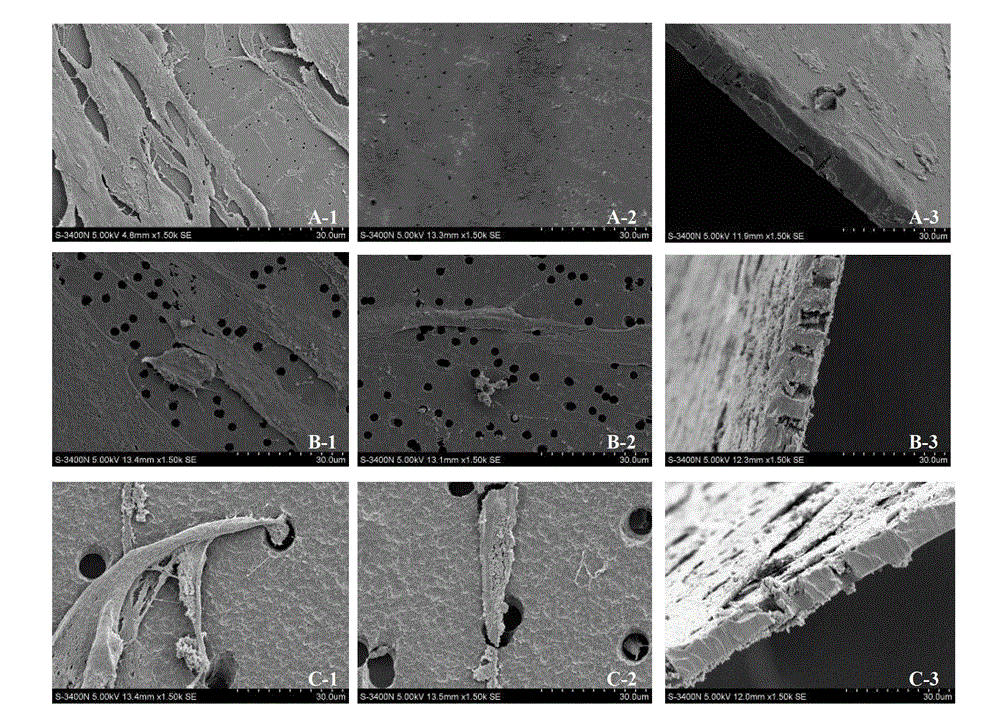
[0032] Example 1. Growth and migration of umbilical cord mesenchymal stem cells on polycarbonate membranes with different pore sizes

[0033] 1. Group processing

[0034] 1. The umbilical cord mesenchymal stem cells were treated with a mesenchymal stem cell medium containing 10 mg / L mitomycin C for 3 hours, and then the cells were washed with the mesenchymal stem cell medium and suspended.

[0035] 2. Group processing:

[0036] The first group: 6 Transwell chambers (polycarbonate membrane at the bottom with a diameter of 24 mm, a pore diameter of 0.4 μm, and a thickness of 10 μm) were placed on a 6-well cell culture plate;

[0037] The second group: 6 Transwell chambers (polycarbonate membrane at the bottom with a diameter of 24 mm, a pore diameter of 1.0 μm, and a thickness of 10 μm) were placed on a 6-well cell culture plate;

[0038] The third group: 6 Transwell chambers (polycarbonate membrane at the bottom with a diameter of 24 mm, a pore diameter of 3.0 μm, and a thick...

Embodiment 2

[0050] Example 2. New Transwell co-culture induces UCMSCs to differentiate into epidermoid cells

[0051] 1. Group processing

[0052] 1. Human epidermal stem cells were treated with epidermal stem cell medium containing 10 mg / L mitomycin C and 10% calf serum for 3 hours, and then cells were washed with epidermal stem cell medium and suspended.

[0053] 2. Treat the umbilical cord mesenchymal stem cells with a mesenchymal stem cell medium containing 10% calf serum for 3 hours, then wash the cells with the mesenchymal stem cell medium and suspend the cells.

[0054] 3. Group processing

[0055] The first group (flow chart see figure 2 A, co-cultivation of the front and back sides of the polycarbonate membrane at the bottom of the Transwell chamber): Under sterile conditions, take 6 Transwell chambers (the diameter of the polycarbonate membrane at the bottom is 24mm, the pore diameter is 0.4μm, and the thickness is 10μm), and the bottom is reversed. In a sterile 15cm petri d...


PUM

PropertyMeasurementUnit
pore sizeaaaaaaaaaa
pore sizeaaaaaaaaaa
thicknessaaaaaaaaaa

Abstract

The invention discloses a method for the directional induction of the differentiation of MSCs. The method for the directional induction of the differentiation of MSCs comprises the following steps: inoculating and adsorbing in-vitro induction cells to the lower surface of a polycarbonate film, inoculating in-vitro MSCs to the upper surface of the polycarbonate film, and co-culturing the induction cells and the MSCs to promote the differentiation of the MSCs to target cells, wherein each of the MSCs and the induction cells cannot pass through the holes of the polycarbonate film. The differentiation of UCMSCs to cuticle cells has a beneficial effect on the clinic skin regeneration research progress in the future. The method develops ideas for the increase of the induction method for the multi-directional differentiation of the MSCs, and provides bases for the construction of tissue engineering skins through adopting the UCMSCs as seed cells.

Description

technical field [0001] The invention relates to a method for directionally inducing differentiation of mesenchymal stem cells. Background technique [0002] Under specific induction conditions in vivo or in vitro, mesenchymal stem cells (MSCs) can not only differentiate into mesoderm mesenchymal tissue cells such as osteoblasts, adipocytes, and muscle cells, but also cross the germ layer boundary and differentiate into ectodermal nerve cells. Cells, glial cells, and endoderm hepatocytes. Therefore, in recent years, MSCs have become a research hotspot in the field of tissue and organ injury repair and regeneration in basic medicine and clinical medicine. The differentiation of MSCs into epidermal cells is one of the key steps in the construction of tissue-engineered skin. [0003] The MSCs differentiation program is a guiding signal that may exist in the microenvironment in which they survive. Through the corresponding signal transduction pathway, the expression of key gene...

Claims


Application Information

Patent Timeline
Patent Type & Authority Applications(China)
IPC IPC(8): C12N5/074
Inventor 柴家科李东杰申传安孙天骏
Owner FIRST HOSPITAL AFFILIATED TO GENERAL HOSPITAL OF PLA
Learn More