
Flow cytometry detection method of shrimp hemocyte mitochondrial membrane potential

A technology of mitochondrial membrane potential and flow cytometry, applied in the field of marine biology, can solve problems such as not being widely used, and achieve the effects of avoiding operation damage, large measurement amount and high accuracy

Inactive Publication Date: 2013-05-29
SOUTH CHINA NORMAL UNIVERSITY
View PDF4 Cites 6 Cited by
  • Summary
  • Abstract
  • Description
  • Claims
  • Application Information

AI Technical Summary

Problems solved by technology

Flow cytometry is now a routine assay of cell function in higher vertebrates, but it has not been widely used in shrimp

Method used


Image

  • Flow cytometry detection method of shrimp hemocyte mitochondrial membrane potential
  • Flow cytometry detection method of shrimp hemocyte mitochondrial membrane potential
  • Flow cytometry detection method of shrimp hemocyte mitochondrial membrane potential

Examples


Embodiment

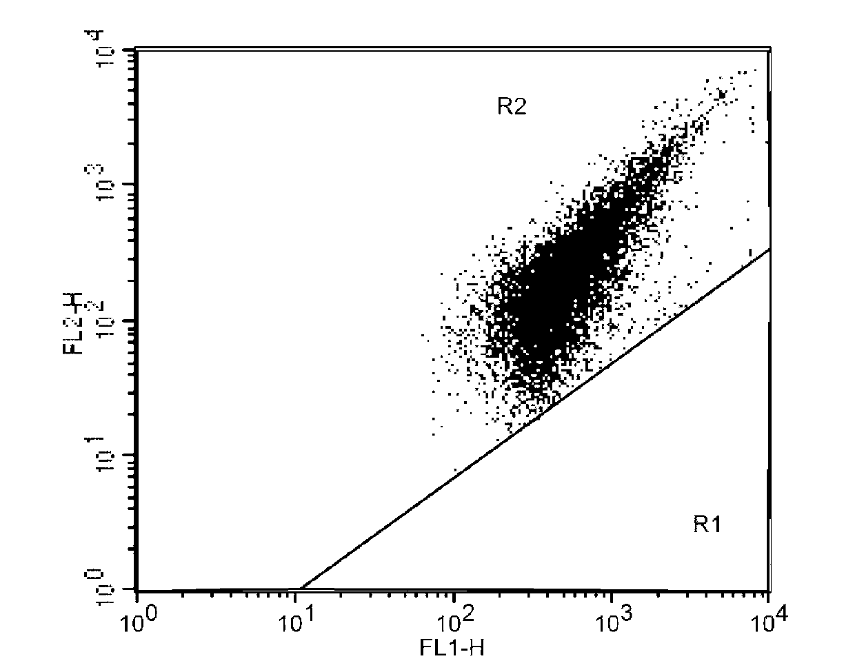
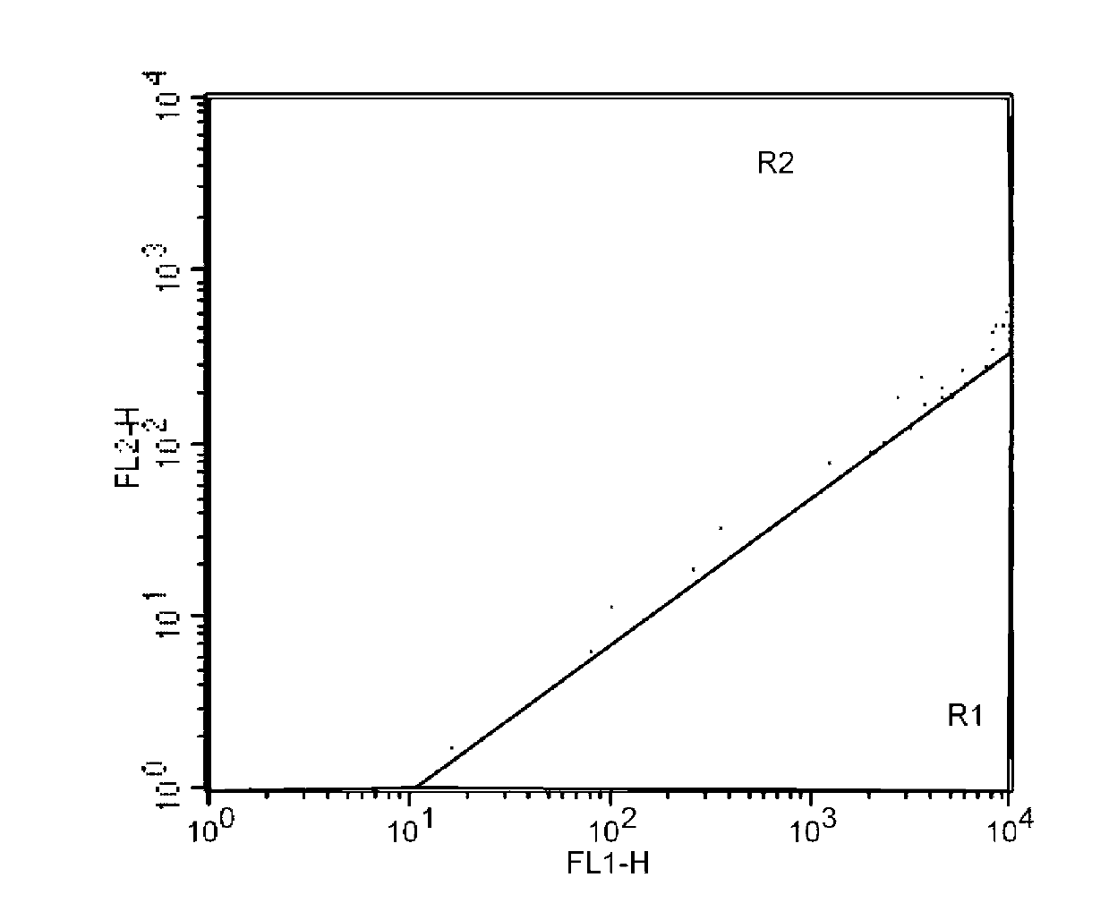
[0027] The mitochondrial membrane potential of isolated hemocytes of Litopenaeus vannamei treated with cadmium ions was measured using a flow cytometry detection method, including the following steps:

[0028] (1) Prepare an anticoagulant suitable for Litopenaeus vannamei: add glucose 20.5g / L, sodium citrate 8g / L, and sodium chloride 4.2g / L to distilled water, adjust the pH to 7.5, autoclave, and cool Store in a 4°C refrigerator for later use.

[0029] (2) Prepare the blood cell suspension of prawns: take out the prawns, wipe off the water on the body surface with cotton balls, and use a sterile syringe with pre-extracted sterilized and pre-cooled anticoagulant to extract and Put the hemolymph with the same volume as the anticoagulant into a sterile centrifuge tube, take enough hemolymph, mix them, and adjust the cell concentration to about 10 with the pre-cooled anticoagulant 6 cells / mL.

[0030] (3) Preparation of negative control group and positive control group: Take 2 t...


PUM


Abstract

The invention discloses a flow cytometry detection method of shrimp hemocyte mitochondrial membrane potential. The method comprises the following steps of preparing shrimp hemocyte suspension; preparing two parts of shrimp cell suspension into a positive control group and a negative control group; adding fluorochrome JC-1 to co-incubate together with hemocyte; determining an area of hemocyte with normal shrimp hemocyte mitochondrial membrane potential and an area of hemocyte with reduced shrimp hemocyte mitochondrial membrane potential by a flow cytometry by the positive control group and the negative control group; carrying out experimental treatment on one part of shrimp hemocyte suspension, or preparing blood hemocyte suspension from shrimps after in vivo experiment treatment as an experiment group; adding the fluorochrome JC-1 to co-incubate together with the hemocyte; and determining the ratio of the hemocyte with reduced shrimp hemocyte mitochondrial membrane potential by the flow cytometry according to the determined area of the hemocyte with normal shrimp hemocyte mitochondrial membrane potential and the area of the hemocyte with reduced shrimp hemocyte mitochondrial membrane potential. The method disclosed by the invention is free of cell washing, prevents the hemocyte from an operation injury, is high in accuracy, good in repeatability, large in determination amount, and simple and rapid to operate; and a method foundation is provided for determination of the shrimp hemocyte mitochondrial membrane potential.

Description

technical field [0001] The invention belongs to the technical field of marine organisms, and in particular relates to a flow cytometry detection method for the mitochondrial membrane potential of shrimp hemocytes. Background technique [0002] Shrimp is an important economic aquatic animal in my country, with a wide variety, mainly including Litopenaeus vannamei, Penaeus monodon, Fenneropenaeus chinensis, Marsupenaeus japonicus, Marsh rosenbergii Shrimp (Macrobrachium rosenbergii), Procambarus clarkii (Procambarus clarkii), etc. Shrimp is an important agricultural product for the adjustment of my country's aquaculture structure, the increase of farmers' production and income, and the country's export earnings. However, in recent years, large-scale morbidity and mortality have frequently occurred in shrimp farming, causing serious economic losses. There are many reasons for this problem, among which a series of adverse effects brought about by the deterioration of the aquacu...

Claims


Application Information

Patent Timeline
Patent Type & Authority Applications(China)
IPC IPC(8): G01N21/64
Inventor 王安利冼健安苗玉涛潘训彬李彬郭慧
Owner SOUTH CHINA NORMAL UNIVERSITY
Learn More