
Wide-field retina microelectrode array

A technology of micro-electrode array and electrode array, which is applied in the field of biomedical signal detection, can solve the problems of inability to obtain higher resolution, high difficulty in processing and implanting retinal prostheses, and achieve the effect of increasing the difficulty of processing

Inactive Publication Date: 2013-02-13
INST OF BIOMEDICAL ENG CHINESE ACAD OF MEDICAL SCI +1
View PDF3 Cites 7 Cited by
  • Summary
  • Abstract
  • Description
  • Claims
  • Application Information

AI Technical Summary

Problems solved by technology

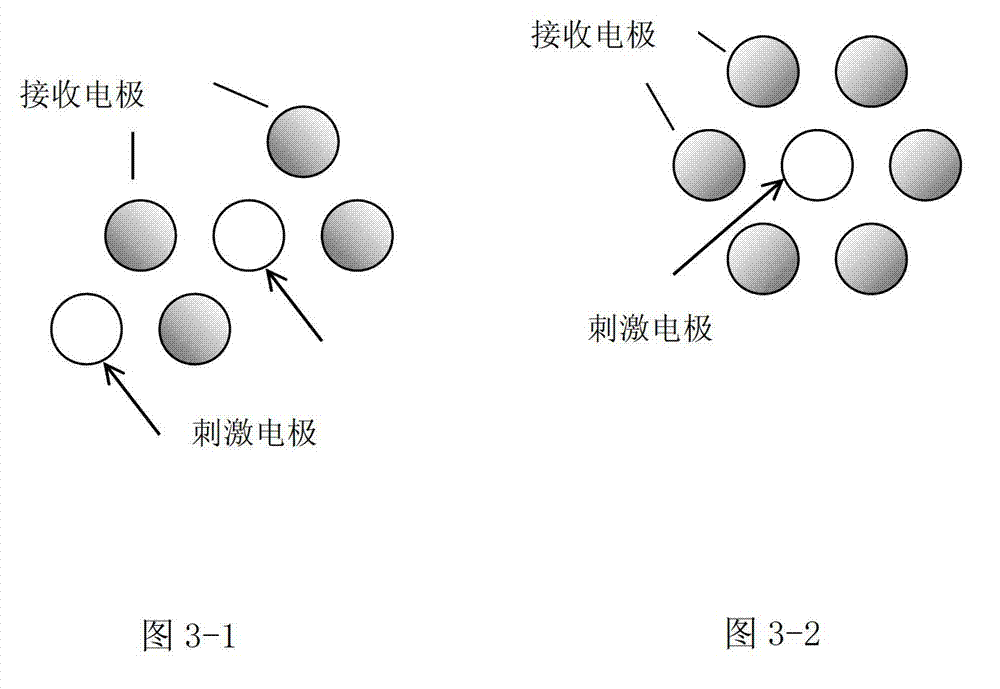
[0005] 2. The electrode array arranged in a hexagonal honeycomb shape is similar to the stimulating electrode array arranged in a square. It is also limited to the field of view in the horizontal direction and cannot obtain higher resolution in the horizontal direction.
[0006] 3. The electrode array of the arc-shaped substrate divides the electrode array substrate according to the natural curvature of the retina. When implanting the retina, it needs to be spliced ​​and fixed to make the synthesized electrode array in a spherical arc shape. Although the array can obtain a wider field of view in the horizontal direction and the vertical direction, the processing and implantation of this retinal prosthesis are too difficult

Method used


Image

  • Wide-field retina microelectrode array
  • Wide-field retina microelectrode array
  • Wide-field retina microelectrode array

Examples


Embodiment Construction

[0027] Embodiments of the present invention are described in further detail below in conjunction with the accompanying drawings:

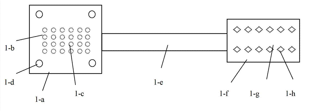
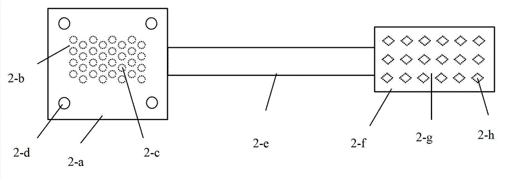
[0028] A wide-field retinal microelectrode array, such as figure 1 and figure 2 As shown, it includes the electrode array part, the electrode array lead-out part and the external electrical stimulation dot matrix part connected in sequence. The electrode array part, the electrode array lead-out part and the external electric stimulation dot matrix part are made integrally, which avoids the complicated process of connection. The thickness is not more than 20μm. The electrode array part, the electrode array lead-out part and the external electrical stimulation dot matrix part are explained separately below:

[0029] exist figure 1 In the shown embodiment, the electrode array part is composed of a biocompatible soft polyester substrate 1-a and a stimulating electrode array 1-b, the stimulating electrode array is embedded on the soft polyester subs...


PUM

PropertyMeasurementUnit
Thicknessaaaaaaaaaa
Lengthaaaaaaaaaa
Lengthaaaaaaaaaa

Abstract

The invention relates to a wide-field retina microelectrode array. The technical scheme includes that an electrode array portion is composed of a soft polyester substrate and a retina stimulating electrode array, the stimulating electrode array is embedded in the soft polyester substrate and is rectangular, the length of the stimulating electrode array in the horizontal direction is longer than that in the vertical direction, a fixing hole is arranged on the soft polyester substrate, a leading-out portion of the electrode array is composed of a soft polyester substrate and an electrode lead which is embedded on the soft polyester substrate, an external electrical stimulation lattice portion is composed of an external electrical stimulation portion substrate and an external stimulation lattice, the external stimulation lattice is embedded on the soft polyester substrate, and the stimulating electrode array is connected with the external stimulation lattice through the electrode lead. According to the wide-field retina microelectrode array, the electrode array which is arranged in a rectangular mode is used, and the length of the stimulating electrode array in the horizontal direction is longer than that in the vertical direction, so that a plurality of cone cells and rod cells can be stimulated in the horizontal direction, the wide view field in the horizontal direction can be provided, and simultaneously, the difficulty in processing of the microelectrode array is not increased.

Description

technical field [0001] The invention belongs to the field of biomedical signal detection, in particular to a wide-view retinal microelectrode array. Background technique [0002] The retina, located on the back wall of the eye, is the light-sensitive area that contains the light-sensitive units rods and cones. The optic nerve carries electrical signals generated by light stimulation to the rods and cones to the brain for visual processing. In the case of retinopathy, age-related macular degeneration (AMD) and retinitis pigmentosa (RP) cone and rod cells degenerate to varying degrees, resulting in vision loss or blindness; however, the optic nerve is not damaged and can still Electrical signals from the retina are transmitted to the brain. Therefore, by giving appropriate stimulation to the part of the retina that has not been damaged, the patient's vision can be improved or restored. The retinal prosthesis (Retinal Prothesis) is aimed at the above-mentioned retinal diseas...

Claims


Application Information

Patent Timeline
IPC IPC(8): A61N1/05A61N1/36
Inventor 谢小波徐圣普汪建涛胡勇崔红岩冯莉
Owner INST OF BIOMEDICAL ENG CHINESE ACAD OF MEDICAL SCI
Learn More