Wide-FOV (field of view) and low-dose Micro-CT (computed tomography) cone beam imaging system

An imaging system and cone-beam technology, applied in the field of high-resolution in vivo scanning imaging, can solve the problems of large radiation dose, small imaging field of view, and changing the biological process of diseases, so as to reduce radiation, ensure imaging quality, and ensure consistency Effect

Inactive Publication Date: 2012-01-18
FOURTH MILITARY MEDICAL UNIVERSITY
View PDF4 Cites 18 Cited by
  • Summary
  • Abstract
  • Description
  • Claims
  • Application Information

AI Technical Summary

Problems solved by technology

[0008] Although the current Mirco-CT has a relatively high resolution, there are still two main problems in live animal imaging. One is the small imaging field of view. At present, the FOV diameter of most systems is less than 80mm, and it is mainly used for ex vivo tissue imaging. Or live imaging of nude mice and rats cannot be used for live imaging of larger animals such as rabbits; second, the radiation dose generated during imaging is relatively large, and experimental animals exposed to high-dose radiation for a long time will change the biological characteristics of the disease. learning processes and creating obvious false impressions
Data show that in adoptive transfer experiments, radiation doses of 1-2 Gy were used to disrupt the mouse immune system, making radiation doses too high for serial imaging in live animals

Method used


Image

  • Wide-FOV (field of view) and low-dose Micro-CT (computed tomography) cone beam imaging system
  • Wide-FOV (field of view) and low-dose Micro-CT (computed tomography) cone beam imaging system
  • Wide-FOV (field of view) and low-dose Micro-CT (computed tomography) cone beam imaging system

Examples


Embodiment Construction

[0037] The technology of the present invention will be described in detail below with reference to the accompanying drawings and specific embodiments.

[0038] 1. System design and composition

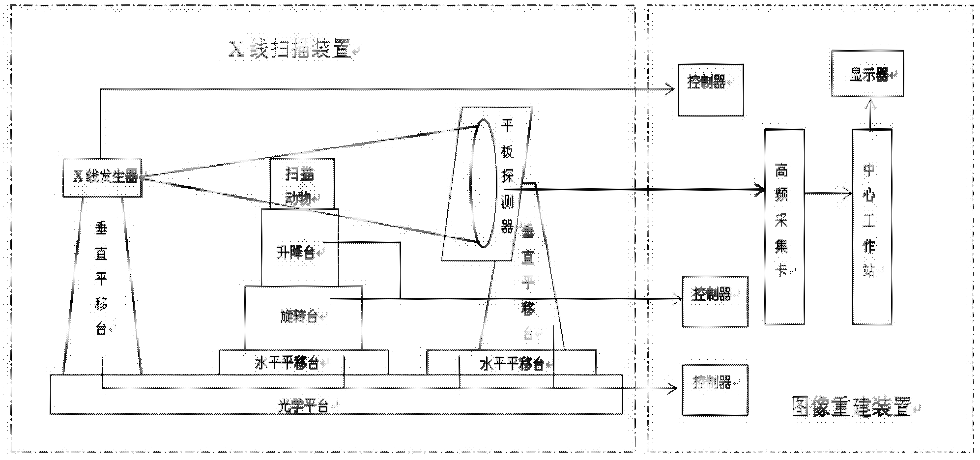
[0039] figure 1 Shown is a schematic structural diagram of the large-field low-dose Mirco-CT cone-beam imaging system. The object to be measured is fixed on the lifting platform, and the X-ray generator and the flat panel detector are respectively fixed on both sides of the object to be measured by two electric translation platforms in the vertical direction, and the directions are opposite. After the X-rays generated by the X-ray generator penetrate the object under test, the remaining X-rays are received by the flat panel detector and converted into projection data of digital signals. The data acquisition card collects these projection data and sends them to the central workstation for 3D reconstruction, and the reconstructed image is displayed on the monitor.

[0040] 2. Design o...


PUM


Abstract

The invention discloses a wide-FOV (field of view) and low-dose Micro-CT (computed tomography) cone beam imaging system based on a flat panel detector. The imaging system mainly comprises a cone beam X-ray scanning device and a three-dimensional image reconstruction device. While the imaging system obtains a resolution ratio (up to 30mu m- 100mu m) satisfying the imaging requirement of a animal tissue structure, the FOV diameter of the imaging system is up to 80mm-150 mm, maximally up to 200mm; and the imaging system can use lower radiation dose (less than 100 mGy), and can realize vivo scan imaging for bigger animal tissue structures, such as a rabbit and the like.

Description

technical field [0001] The invention is a small animal Mirco-CT cone-beam tomography system based on a flat panel detector, which realizes high-resolution live scanning imaging of larger animals such as rabbits by combining low-dose and large-field imaging. Background technique [0002] Computed tomography (CT) obtains the X-ray projections of the cross-section of the object in different directions, and uses a computer to reconstruct the cross-sectional image of the object to obtain a three-dimensional image of the internal structure of the object. Since Hounsfield invented the world's first CT equipment in 1967, this technology has been widely used in various fields such as medicine, pharmacy, materials science, industry, agriculture, engineering and archaeology. [0003] Although the spatial resolution of clinical CT images has reached the order of submillimeters, it is difficult to meet the requirements for ultra-high structural resolution in basic medical research and ma...

Claims


Application Information

Patent Timeline
Patent Type & Authority Applications(China)
IPC IPC(8): A61B6/03
CPCA61B6/508
Inventor 廖琪梅卢虹冰刘文磊刘洋
Owner FOURTH MILITARY MEDICAL UNIVERSITY
Why Eureka
Try Eureka