
Dual-channel whole-eye optical coherence tomography system and imaging method

An optical coherence tomography and imaging system technology, applied in the field of laser medical equipment and laser imaging, can solve problems such as OCT systems that have not yet been discovered, and achieve the effect of saving procedures and costs, and having a simple and compact system structure

Active Publication Date: 2011-12-21
GUANGDONG FORTUNE NEWVISION TECH
View PDF8 Cites 20 Cited by
  • Summary
  • Abstract
  • Description
  • Claims
  • Application Information

AI Technical Summary

Problems solved by technology

[0003] To date, no OCT system capable of simultaneously imaging the anterior segment and fundus has been found in the prior art and equipment

Method used


Image

  • Dual-channel whole-eye optical coherence tomography system and imaging method
  • Dual-channel whole-eye optical coherence tomography system and imaging method
  • Dual-channel whole-eye optical coherence tomography system and imaging method

Examples


Embodiment Construction

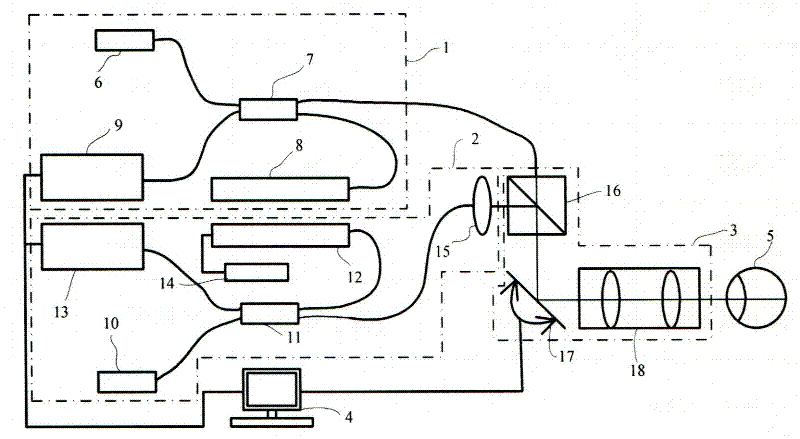
[0041] figure 2 The dual-channel whole-eye optical coherence tomography system of the present invention is shown, including a fundus optical coherence tomography part 1 , an anterior segment optical coherence tomography part 2 , a detection arm 3 and a PC control platform 4 .

[0042] The fundus optical coherence tomography part 1 includes a first near-infrared light source 6, a first fiber coupler (2*2 fiber coupler) 7, a first reference arm 8, and a first spectrometer 9. The first near-infrared The light beam emitted by the infrared light source 6 is input into the first fiber coupler 7, and then output to the detection arm 3 and the first reference arm 8 respectively, and the fundus backscattered light signal returned by the detection arm 3 and the first reference The specularly reflected optical signal returned by the arm 8 is output to the first spectrometer 9 after being interfered by the first optical fiber coupler 7, and the first spectrometer 9 is connected to the PC...


PUM


Abstract

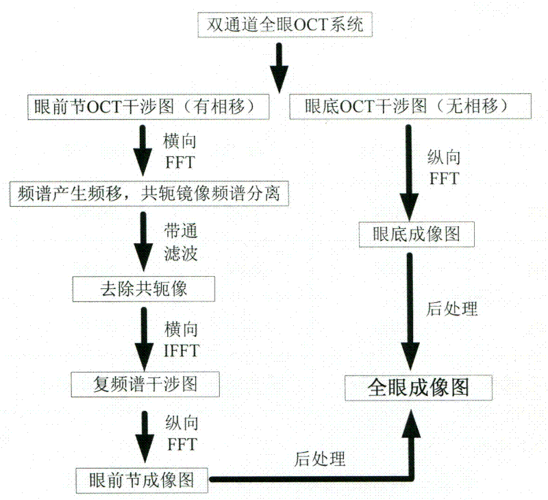
The invention discloses a dual-channel whole-eye OCT system, which includes a fundus OCT part, an anterior segment OCT part, a detection arm and a PC control platform; the light beam emitted by the near-infrared light source of the fundus part is output to the detection arm and the first reference through a fiber coupler. arm, the returned optical signal forms interference and then outputs to the first spectrometer; the light beam emitted by the near-infrared light source of the anterior segment is output to the detection arm and the second reference arm through the fiber coupler, and the returned optical signal forms interference and is output to the second spectrometer. The present invention also provides a method for performing whole-eye OCT by using the above-mentioned imaging system, in which the fundus tomographic image and the anterior segment tomographic image are post-processed through a PC control platform, thereby combining to obtain a whole-eye optical coherence tomographic image. The structure and method of the present invention are ingenious, so that the fundus part and the anterior segment part are imaged at the same time, and the OCT imaging of the whole eye is realized. The system structure is simple and compact, and the ophthalmic examination procedure and cost are greatly saved.

Description

technical field [0001] The present invention relates to a laser medical device and a laser imaging method, in particular to a laser ophthalmology examination device and a dual-channel all-eye optical coherence tomography method, specifically, a dual-channel all-eye optical coherence tomography that simultaneously realizes anterior segment and fundus imaging Coherent tomography imaging system and imaging method. Background technique [0002] During ophthalmological examination, the imaging examination of the anterior segment and fundus is very important. It is necessary to detect the anterior segment and fundus before various surgical treatments such as excimer laser surgery. In many patients with eye diseases such as diabetic retinopathy and macular It is also an indispensable item when checking for sex change, etc. In the past ten years, the research of Optical Coherence Tomography (OCT) has made important progress, especially in ophthalmology. widely used. However, curr...

Claims


Application Information

Patent Timeline
IPC IPC(8): A61B3/12A61B3/14
Inventor 周传清王纬超
Owner GUANGDONG FORTUNE NEWVISION TECH
Learn More