
Method for calculating brain blood volume on basis of stable status method

A technology of cerebral blood volume and steady state method, which is applied in computer tomography scanners, diagnostic recording/measurement, medical science, etc., can solve the problems of difficult to achieve three-dimensional perfusion imaging, easy missed diagnosis of lesions, etc., to reduce dependence and sensitivity. degree, the effect of reducing radiation dose

Inactive Publication Date: 2011-07-13
姜卫剑 +1
View PDF0 Cites 12 Cited by
  • Summary
  • Abstract
  • Description
  • Claims
  • Application Information

AI Technical Summary

Problems solved by technology

But so far, there are still many problems that have not been completely resolved. For example, most of the current researches use a layer with a thickness of 10mm as the research object (including the use of multi-slice spiral CT). Even if 64-slice spiral CT is used, only 40mm scanning can be performed, so lesions located outside the scanning layer are easily missed, and three-dimensional perfusion imaging of the entire brain tissue is currently difficult to achieve

Method used


Image

  • Method for calculating brain blood volume on basis of stable status method
  • Method for calculating brain blood volume on basis of stable status method
  • Method for calculating brain blood volume on basis of stable status method

Examples


Embodiment 1

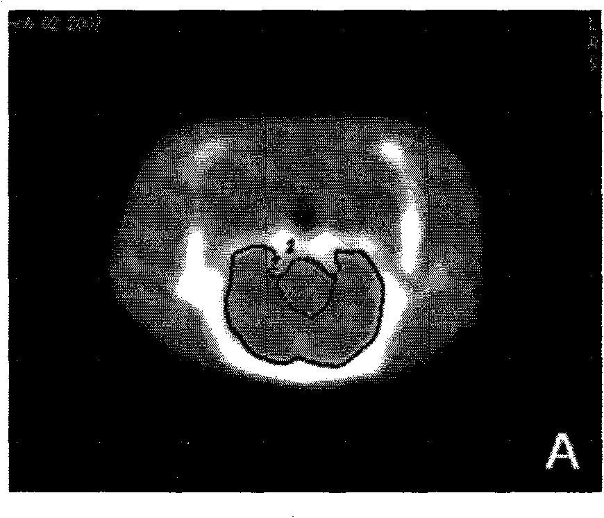
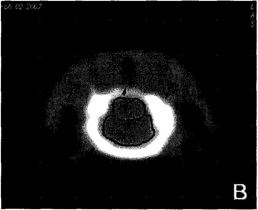
[0043]The study was approved by the local Animal Care Committee. Eleven healthy rats were included in the feasibility study. Continuous CT acquisition was performed on the head of the rat first, and then continuous CT acquisition was performed on the same position of the head of the rat after intravenous injection of contrast agent. We measured the CBV values ​​of the parieto-occipital lobe, deep brain gray matter, cerebellum and brainstem with steady-state and dynamic methods, respectively. The results were: the CBV values ​​of the parieto-occipital lobe, deep gray matter, cerebellum and brainstem measured by the steady-state method were 3.19±0.83mL / 100g, 3.22±0.82mL / 100g, 4.54±1.11mL / 100g, and 3.87± 1.00mL / 100g. The average CBV values ​​of the parieto-occipital lobe, deep gray matter, cerebellum and brainstem of the control group were measured by dynamic method were 3.30±0.69mL / 100g, 3.55±0.87mL / 100g, 4.56±0.93mL / 100g, and 3.97±0.81 mL / 100g. The consistency of CBV values...


PUM


Abstract

The invention relates to a method for calculating brain blood volume. The method is used for calculating the brain blood volume on the stable status CT (Computed Tomography) brain perfusion imaging principle. The method comprises the following steps of (1) carrying out common whole brain CT scanning to obtain a brain tissue structure image before contrast agent injection; injecting the contrast agent to the vessel exciting area to reach the stable status, and obtaining the brain tissue structure image of the same position; (3) deleting an overhigh brain blood volume value with a vessel pixel deleting method, and deleting the interference of the artery in the existing area to the brain blood content measuring value; (4) and calculating the brain blood flow value of the brain tissue by calculating the ratio of the brain tissue to the signal change in the vessel according to a formula. With the method, all brain scanning can be carried out and the radial dosage of the perfusion scanning can be reduced, thus the method for calculating brain blood volume can be widely applied into the clinical trial.

Description

Background technique [0001] Acute cerebrovascular disease is a common disease of the nervous system, and its morbidity and mortality tend to increase year by year, of which about 67% to 80% are acute ischemic cerebrovascular disease (AICVD). In order to timely screen AICVD patients within the treatment time window and formulate appropriate treatment plans, clinicians need to obtain the status of cerebral blood flow of patients. Positron emission computed tomography (PET), single photon emission computed tomography (SPECT), xenon-CT (Xe-CT) and other imaging techniques can provide a lot of useful information about the pathophysiological changes of brain tissue in AICVD patients. However, there are some deficiencies in these inspection methods, which limit the application and promotion. The current calculation of cerebral blood volume based on computed tomography (CT) images usually adopts a fast dynamic multi-temporal acquisition method to collect data, and then calculates the...

Claims


Application Information

Patent Timeline
Patent Type & Authority Applications(China)
IPC IPC(8): A61B5/0275A61B6/03
CPCA61B6/507
Inventor 姜卫剑申皓马宁娄昕
Owner 姜卫剑
Learn More