
Digital imaging system of urology surgery

A urology, digital imaging technology, applied in the field of urology digital imaging system, can solve the problems of inability to share image information, increase treatment costs, and isolate patient information.

Active Publication Date: 2010-10-27
SHENZHEN HUIKANG SOFTWARE TECH
View PDF5 Cites 17 Cited by
  • Summary
  • Abstract
  • Description
  • Claims
  • Application Information

AI Technical Summary

Problems solved by technology

The traditional process wastes the time of doctors and patients waiting for DR films and repeatedly entering patient information; printing films also wastes resources and increases treatment costs; the complexity of the process also reduces the efficiency of treatment
Therefore, the current status of urology is obviously not in line with the development trend of PACS. The isolation of patient information and the inability to share image information have greatly hindered the development of urology.

Method used


Image

  • Digital imaging system of urology surgery
  • Digital imaging system of urology surgery
  • Digital imaging system of urology surgery

Examples


Embodiment Construction

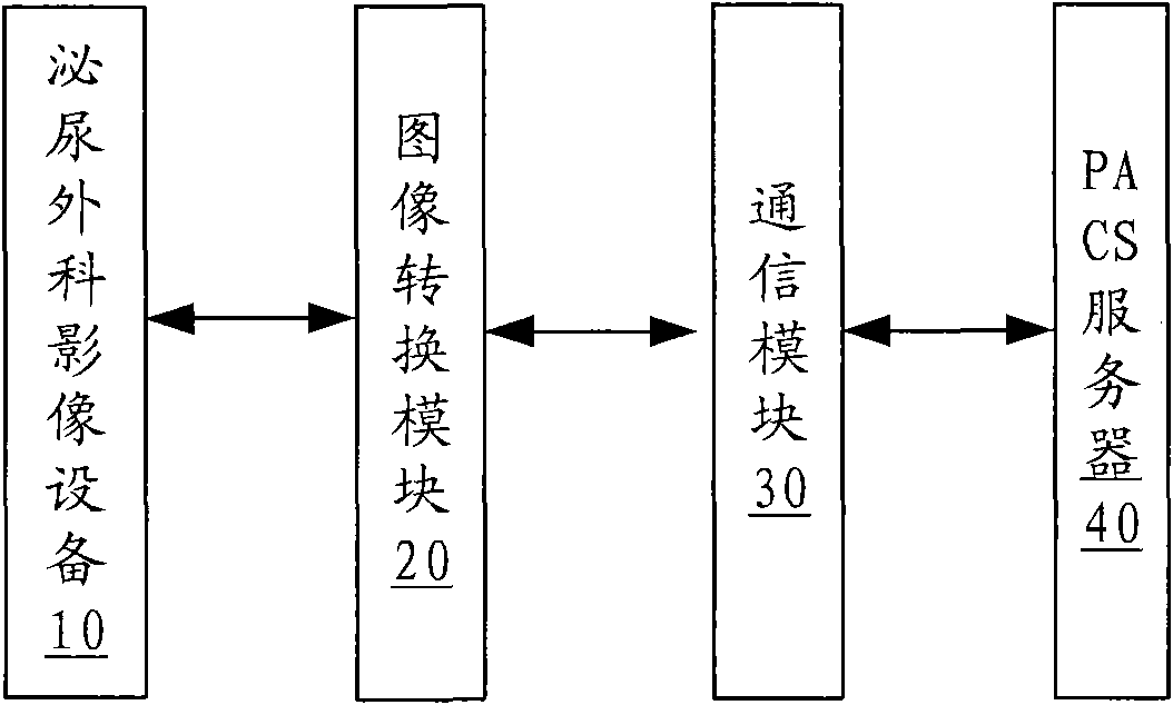
[0015] Such as figure 1 As shown, a urology digital imaging system includes a urology imaging device 10, an image conversion module 20, a communication module 30 and a PACS server 40, wherein:

[0016] The urology imaging device 10 is used for collecting urology images of patients. The urology imaging device 10 can be an extracorporeal lithotripter, a urology operating bed, an ultrasonic diagnostic instrument, an endoscope inspection machine, etc. When a patient goes to the urology department for treatment, images can be collected through these imaging devices.

[0017] The image conversion module 20 is used for converting the urology surgery image into a DICOM standard image of the urology surgery, and writing the patient's medical information into the DICOM standard image. Since the images collected by the urology imaging device 10 are all non-DICOM standard images, usually in BMP format, the non-DICOM standard images cannot be shared in the PACS server, so it is necessary ...


PUM


Abstract

The invention provides a digital imaging system of the urology surgery, comprising an imaging device of the urology surgery, an image conversion module, a communication module and a PACS server, wherein the imaging device of the urology surgery is used for collecting images of urology surgery of patients; the image conversion module is used for converting the images of urology surgery into DICOM standard images of urology surgery and writing medical treatment information of patients in the DICOM standard images; the communication module is used for sending the DICOM standard image of urology surgery and medical treatment information of patients to a PACS server; and the PACS server is used for receiving the DICOM standard images of urology surgery and medical information of patients for storage. By adopting the invention, digitization of imaging device of the urology surgery and sharing of the image information of the urology surgery can be realized.

Description

【Technical field】 [0001] The invention relates to the technical field of medical equipment, in particular to a digital imaging system for urology. 【Background technique】 [0002] Imaging equipment in the hospital's urology department mainly includes extracorporeal lithotripters, urology operating beds, ultrasonic diagnostic equipment, and endoscopic inspection machines. At present, urology imaging equipment basically has an analog output interface, and cannot be connected to PACS (Picture Archiving and Communication Systems, image archiving and communication system), because PACS only connects with digital DICOM (Digital imaging and Comunicaitons in Medicine, digital imaging and communication Standard) interface imaging equipment, so PACS basically ignores urology. [0003] At present, monitors are still used to observe images by imaging equipment in the Department of Urology, and many of the configured workstations only record medical records. Taking the lithotripsy machi...

Claims


Application Information

Patent Timeline
Patent Type & Authority Applications(China)
IPC IPC(8): A61B5/00
Inventor 蔡虹陈明
Owner SHENZHEN HUIKANG SOFTWARE TECH
Learn More