Quick partition method of three-dimensional medical image on basis of video card parallel computing

A medical image and parallel computing technology, which is applied in the field of medical image processing and medical image segmentation, can solve the problems of not considering the original image to save storage space and not applicable to applications, etc.

Inactive Publication Date: 2010-07-21
TSINGHUA UNIV
View PDF2 Cites 7 Cited by
  • Summary
  • Abstract
  • Description
  • Claims
  • Application Information

AI Technical Summary

Problems solved by technology

In the paper "OnImplementing Graph Cuts on CUDA" published by Hussein et al. in 2007 and the paper "CUDA Cuts: FastGraph Cuts on the GPU" published by Vineet et al. in 2008, they both used this architecture to achieve two-dimensional The CUDA-style Graph-Cuts method of the image, however, they do not consider the problem of saving storage space when the original image is large in size, so they are not suitable for applications in three-dimensional spaces that require more stringent storage space

Method used


Image

  • Quick partition method of three-dimensional medical image on basis of video card parallel computing
  • Quick partition method of three-dimensional medical image on basis of video card parallel computing
  • Quick partition method of three-dimensional medical image on basis of video card parallel computing

Examples


Embodiment Construction

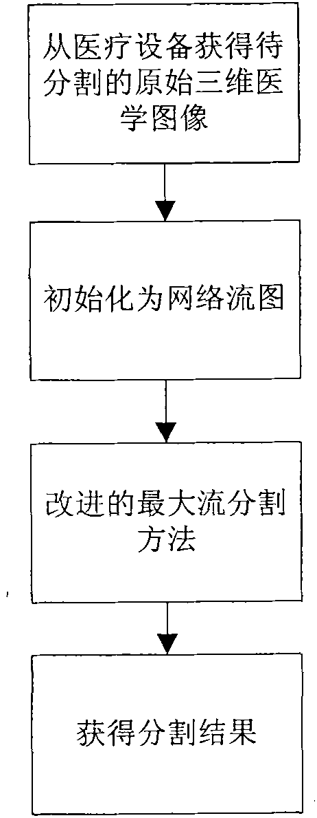
[0030] The present invention proposes a method for fast segmentation of three-dimensional medical images based on graphics card parallel computing, which is characterized in that the method adopts the Graph-Cuts method accelerated based on CUDA technology to segment the original three-dimensional medical images (from medical equipment (such as CT machines) or MRI machine) for segmentation processing; such as figure 1 shown, including the following steps:

[0031] 1) Initialize the original 3D medical image to be segmented as a network flow graph;

[0032] 2) The improved maximum flow method is used for segmentation processing on the network flow graph, and the segmentation result of the 3D medical image is obtained;

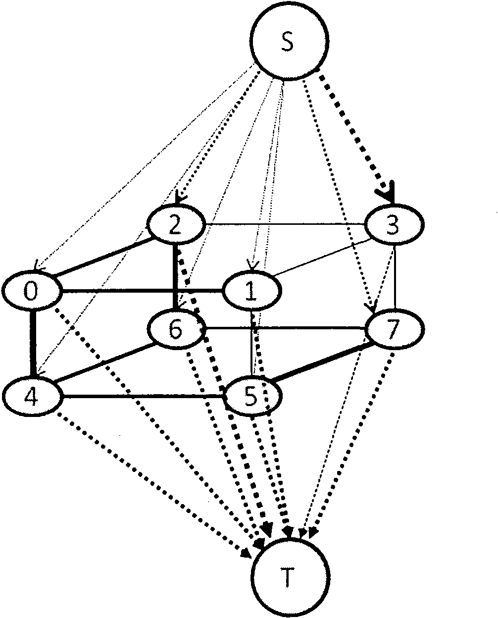
[0033] The above steps 1) initialize the original 3D medical image to be segmented into a network flow graph, such as figure 2 As shown, it specifically includes the following steps:

[0034] 11) First construct the basic structure of the network flow graph c...


PUM


Abstract

The invention relates to a quick partition method of a three-dimensional medical image on the basis of video card parallel computing, belonging to the filed of medical image partitioning. In the method, firstly the basic framework of a network flow graph, which consists of nodes, end points and weighted edges, is built according to an original three-dimensional medical image; then, the logic structure of the network flow graph is built: the storage structure of the network flow graph is built so as to obtain the network flow graph which finishes practical storage; two stages of parallel relabel operation and push operation which are realized on the basis of CUDA is carried out on the network flow graph which finishes practical storage; when the excess flow value of an active node is updated to zero, the active node becomes a common node, and maximal flow from S to T can be obtained from the network flow graph; and when the capacity value in the minimum partitioning corresponding to the maximal flow is equal to the edge of the flow value, a boundary for partitioning a target organ and the background can be formed to obtain a partition result. The method of the invention can greatly improve the operating speed of the partition method in the premise of keeping high processing precision.

Description

technical field [0001] The invention belongs to the technical fields of medical image processing and medical image segmentation, and in particular relates to a fast segmentation method of a three-dimensional medical image based on graphics card parallel computing. Background technique [0002] In visualized medical operations, the three-dimensional configuration technology of human organs and lesions brings great convenience to doctors, and the premise of three-dimensional configuration is the original three-dimensional image composed of a large number of two-dimensional medical image slices. (such as CT, MRI images) to accurately segment the target organ or tumor image. This segmentation has always been a very difficult problem in the field of medical image processing, especially under the premise of both the accuracy of segmentation and the speed of processing. [0003] In the past, images of various organs were extracted from medical images by manually delineating the bo...

Claims


Application Information

Patent Timeline
Patent Type & Authority Applications(China)
IPC IPC(8): G06T17/00G06T7/00
Inventor 王宏杨凡翟伟明宋亦旭赵雁南贾培发
Owner TSINGHUA UNIV
Why Eureka
Try Eureka