
Fusion protein and use of fusion protein in detection of histoplasma capsulatum

A technology of histoplasma bacteria and fusion protein is applied in the field of enzyme-linked immunosorbent assay to detect capsular histoplasma bacteria, which can solve the problem that large-scale promotion in primary medical institutions cannot be achieved, and achieves simple and easy preparation method, accurate use, and high efficiency. The effect of low production cost

Inactive Publication Date: 2011-05-25
CENT SOUTH UNIV
View PDF0 Cites 0 Cited by
  • Summary
  • Abstract
  • Description
  • Claims
  • Application Information

AI Technical Summary

Problems solved by technology

This method has good sensitivity and high specificity, but due to the limitation of instruments, this method cannot be widely promoted in primary medical institutions

Method used


Image

  • Fusion protein and use of fusion protein in detection of histoplasma capsulatum
  • Fusion protein and use of fusion protein in detection of histoplasma capsulatum
  • Fusion protein and use of fusion protein in detection of histoplasma capsulatum

Examples


Embodiment Construction

[0033] The present invention will be described in further detail below with specific examples.

[0034] 1. Cloning of the M gene fragment of Histoplasma capsulatum

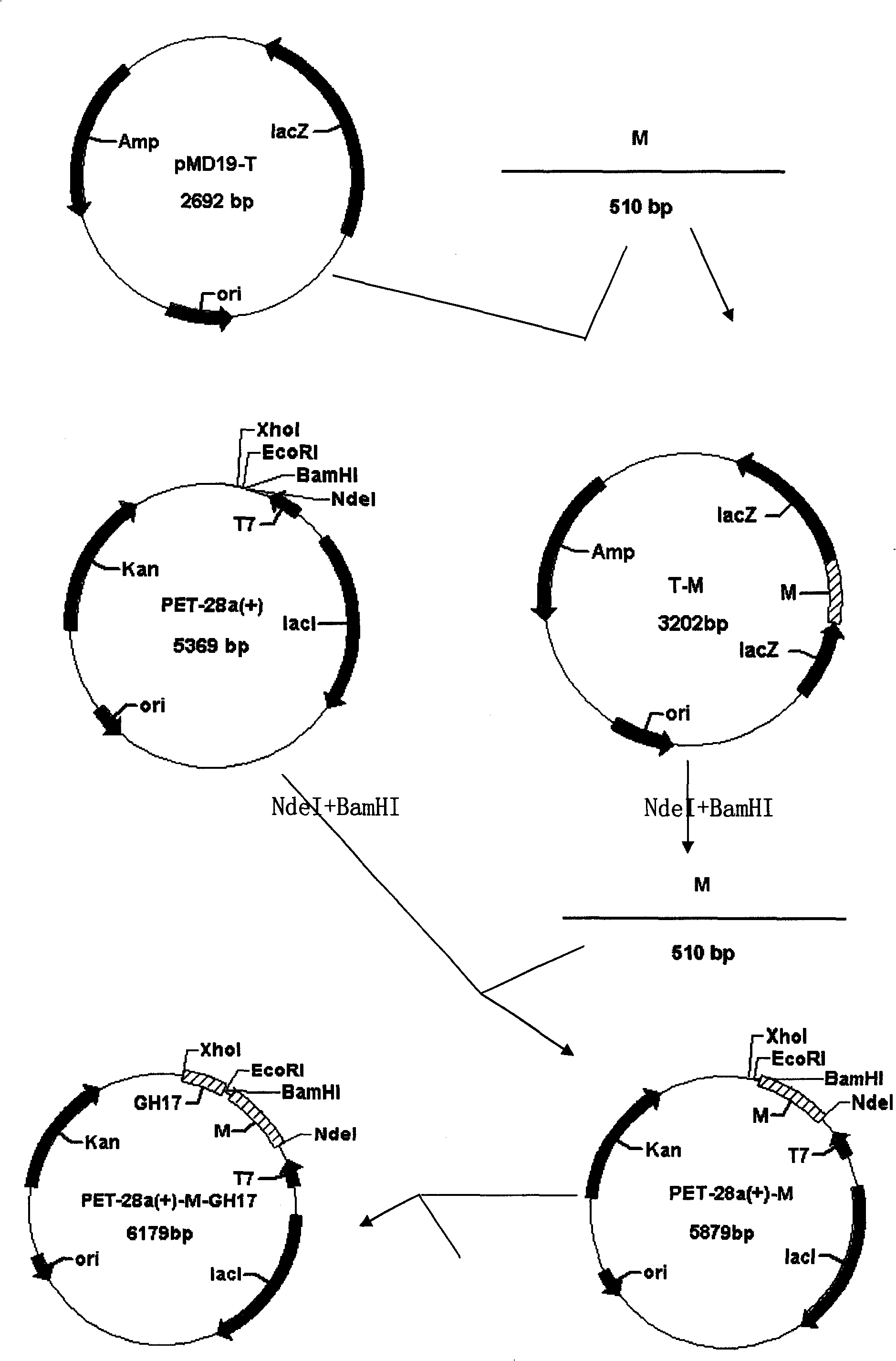
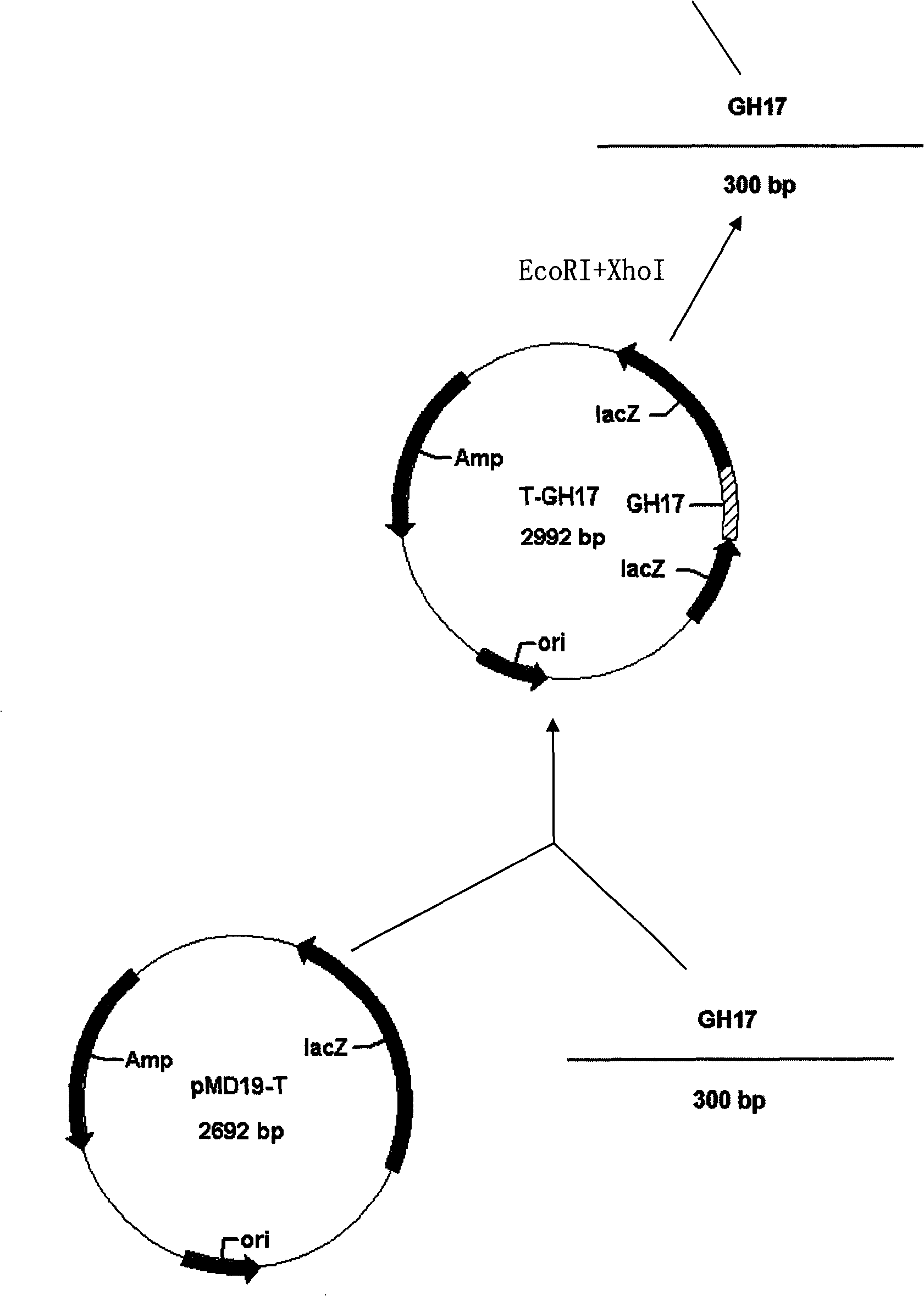
[0035] as attached figure 1 As shown, Histoplasma capsulatum was inoculated on the slant medium of glucose agar, cultured at 25°C for 3 weeks, and its genomic DNA was collected and extracted. Using the extracted Histoplasma capsulatum genomic DNA as a template, the M gene fragment of Histoplasma capsulatum was obtained by PCR method, and the primer sequences were as follows:

[0036] F1: 5'GCCATATGCCTCTAAACACGGCCGC 3' (upstream primer)

[0037] R1: 5'GCGGATCCTTTGTTGTTGGCGCTGTTAA 3' (downstream primer)

[0038] The PCR reaction conditions were denaturation at 94°C for 1 minute, annealing at 55°C for 15 seconds, extension at 72°C for 40 seconds, a total of 30 cycles, and finally extension at 72°C for 5 minutes. The PCR product was subjected to 1% agarose gel electrophoresis, recovered by cutting the gel and conn...


PUM


Abstract

The invention discloses a fusion protein, an ELISA kit containing the fusion protein and the use of the fusion protein in the detection of histoplasma capsulatum. A DNA of a separated genome of the hiThe invention discloses a fusion protein, an ELISA kit containing the fusion protein and the use of the fusion protein in the detection of histoplasma capsulatum. A DNA of a separated genome of the histoplasma capsulatum is used as a template, and an M gene fragment and a GH17 fragment are obtained through the amplification of a PCR method; the M gene fragment and the GH17 fragment of the histoplastoplasma capsulatum is used as a template, and an M gene fragment and a GH17 fragment are obtained through the amplification of a PCR method; the M gene fragment and the GH17 fragment of the histoplasma capsulatum are inserted into a prokaryotic expression vector from a specific direction to build a M-GH17 gene fragment recombined prokaryotic expression vector; the M-GH17 recombined prokaryotic esma capsulatum are inserted into a prokaryotic expression vector from a specific direction to build a M-GH17 gene fragment recombined prokaryotic expression vector; the M-GH17 recombined prokaryotic expression vector is transferred to a host cell, and a fusion protein M-GH17 is obtained through screening, recombination, monocoloning, induction and specific expression; and the fusion protein M-GH17xpression vector is transferred to a host cell, and a fusion protein M-GH17 is obtained through screening, recombination, monocoloning, induction and specific expression; and the fusion protein M-GH17 is further used to prepare the ELLSA kit for detecting the histoplasma capsulatum. The ELISA kit containing the fusion protein M-GH17 is used in the detection of blood serum to be detected accordingis further used to prepare the ELLSA kit for detecting the histoplasma capsulatum. The ELISA kit containing the fusion protein M-GH17 is used in the detection of blood serum to be detected accordingto a common ELISA method and can detect the histoplasma capsulatum of a sample of the blood serum to be detected by determining male gender, female gender and suspicious judgment critical value accordto a common ELISA method and can detect the histoplasma capsulatum of a sample of the blood serum to be detected by determining male gender, female gender and suspicious judgment critical value according to a light absorption value after termination.ing to a light absorption value after termination.

Description

technical field [0001] The invention relates to the detection of fusion protein and capsular histoplasma, in particular to a method for detecting capsular histoplasma by enzyme-linked immunosorbent assay. Background technique [0002] Capsular histoplasmosis (Histoplasmosis, HP) is caused by the biphasic fungus Histoplasma capsulatum (Histoplasma capsulatum, HC). Since the first case of HP was reported in my country in 1958, there have been clinical reports of the disease one after another. However, because the clinical symptoms of capsular histoplasmosis are similar to those of pulmonary tuberculosis and kala-azar; the pathological forms are easily confused with Penicillium marneffei, Leishmania donovani, Toxoplasma gondii, Cryptococcus neoformans, etc. The understanding of the disease among medical workers is generally insufficient, so there is a high rate of missed diagnosis and high rate of misdiagnosis. Disseminated HP often dies due to failure to receive timely and co...

Claims


Application Information

Patent Timeline
Patent Type & Authority Patents(China)
IPC IPC(8): C12N15/62C12N15/70G01N33/569C12P21/02C12R1/19G01N33/86C12N1/21C07K19/00
Inventor 冯俊涛余铭恩李晓照胡成平顾其华
Owner CENT SOUTH UNIV
Learn More