X-ray computer tomography device and tomography method

A technology of tomography and X-rays, applied in computer tomography scanners, instruments for radiological diagnosis, diagnosis, etc., can solve problems such as increased X-ray exposure, unnecessary exposure of X-rays, and changing shooting schedules

Active Publication Date: 2009-03-04
TOSHIBA MEDICAL SYST CORP
View PDF2 Cites 12 Cited by
  • Summary
  • Abstract
  • Description
  • Claims
  • Application Information

AI Technical Summary

Problems solved by technology

That is, the above-mentioned prior art can acquire a required amount of X-ray intensity distribution data of a predetermined specific phase even if an abnormal heartbeat occurs before imaging, but since the imaging cannot be changed according to the abnormal heartbeat before imaging, schedule, so there is the issue of unnecessary exposures that generate x-rays
[0013] Here, it is described that in the retrospective electrocardiogram gating method, in order to obtain the necessary amount of X-ray intensity distribution data of a predetermined specific phase, there is a problem that the X-ray exposure amount becomes large, but there is also a problem in the prospective electrocardiogram gating method. same question

Method used


Image

  • X-ray computer tomography device and tomography method
  • X-ray computer tomography device and tomography method
  • X-ray computer tomography device and tomography method

Examples


Embodiment 1

[0027] [Summary and Features of the X-ray CT Apparatus of Embodiment 1]

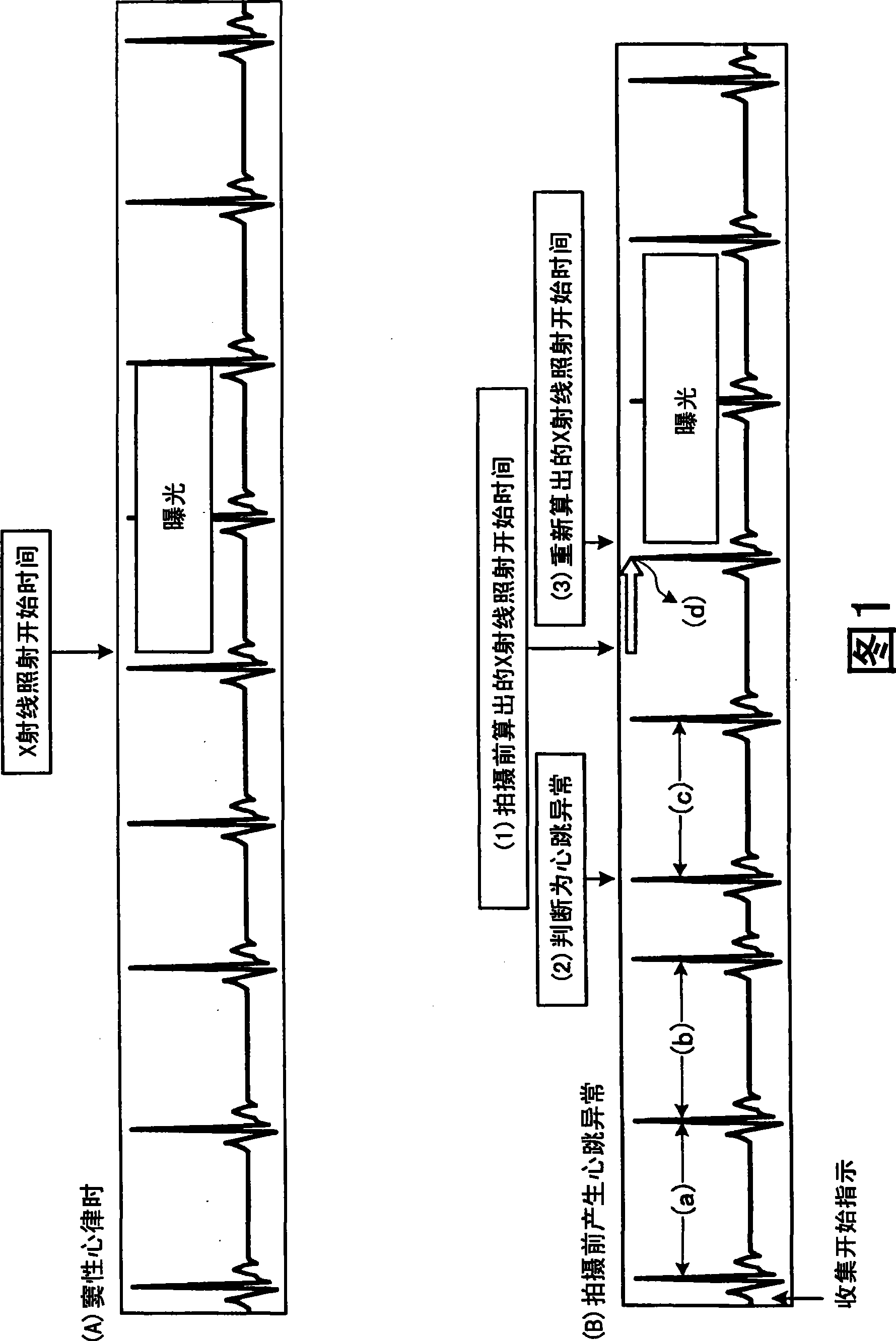
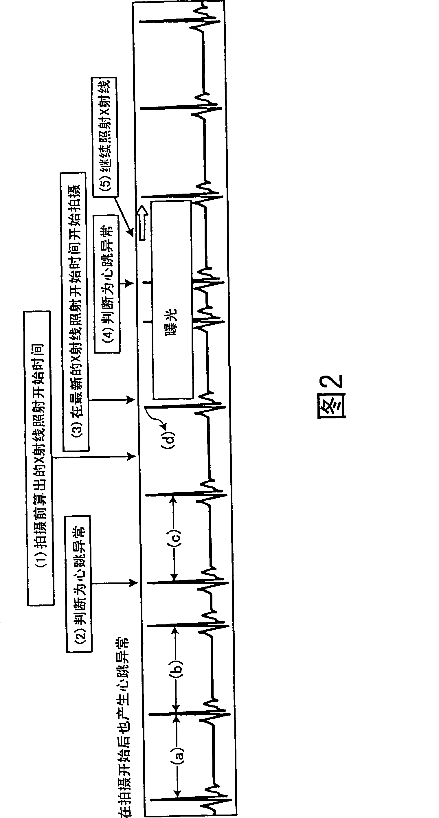
[0028] First, main features of the X-ray CT apparatus of Embodiment 1 will be specifically described using FIG. 1 and FIG. 2 . FIG. 1 is a diagram for explaining the outline and characteristics of an X-ray CT apparatus according to Embodiment 1, and FIG. 2 is a diagram for describing characteristics of the X-ray CT apparatus according to Embodiment 1. As shown in FIG.

[0029] The gist of the X-ray CT apparatus in Embodiment 1 is to irradiate X-rays to a subject equipped with an electrocardiograph, detect the X-rays transmitted through the subject, and obtain the The tomography for reconstructing the tomographic image of the heart in the subject is started based on the X-ray intensity distribution data that is information of the detected X-rays at the start time of imaging.

[0030]That is, the X-ray CT apparatus in Example 1 performs tomography for reconstructing a tomographic image of the heart in the...


PUM


Abstract

An X-ray CT apparatus detects a predetermined characteristic wave from each electrocardiographic waveform acquired from an electrocardiograph, predicts intervals with which the characteristic wave appears based on appearance times of the detected predetermined characteristic wave in each electrocardiographic waveform, and determines an X-ray irradiation start time based on the predicted interval time. After an instruction to start collection of X-ray intensity distribution data is received, if an appearance interval of a detected predetermined characteristic wave is within an acceptable normal-heartbeat range, it is determined as a normal heartbeat. If the appearance interval of the detected predetermined characteristic wave is outside the acceptable normal-heartbeat range, it is determined as an abnormal heartbeat. During a period after receiving the instruction to start the collection until reaching a time of starting the collection of X-ray detection information, when determined as an abnormal heartbeat, an X-ray irradiation start time is recalculated, and then determined.

Description

[0001] References to related applications [0002] This application enjoys the priority right of the Japanese patent application No. 2007-223035 filed on August 29, 2007, and the entire content of the Japanese patent application is cited in this application. technical field [0003] The invention relates to an X-ray computed tomography apparatus and a tomography method. Background technique [0004] In the prior art, an X-ray computed tomography apparatus (hereinafter referred to as an X-ray CT apparatus, CT: Computed Tomography) reconstructs the Provide a tomographic image representing the shape information of the human body tissue in the subject on the plane (tomographic plane) irradiated with X-rays, so that in various medical activities based on disease diagnosis, treatment, operation planning, etc. , plays an important role. [0005] In cardiac examinations for diagnosing heart diseases and the like using cardiac tomographic images of a subject provided by an X-ray CT...

Claims


Application Information

Patent Timeline
Patent Type & Authority Applications(China)
IPC IPC(8): A61B6/03A61B5/0402
CPCA61B6/503A61B6/032A61B5/0456A61B5/352
Inventor 椋本豪神长茂生安田寿山鼻将央森野克人瀬户博光
Owner TOSHIBA MEDICAL SYST CORP
Why Eureka
Try Eureka