Vaccine conjugate including a human chorionic gonadotropin beta subunit antigen linked to an anti-mannose receptor (mr) antibody
A conjugate and antibody technology, applied in the direction of antibody mimic/scaffold, anti-receptor/cell surface antigen/cell surface determinant immunoglobulin, for targeting specific cell fusion, etc.
- Summary
- Abstract
- Description
- Claims
- Application Information
AI Technical Summary
Problems solved by technology
Method used
Image
Examples
Embodiment 1
[0245] Example 1: Production of βhCG-B11
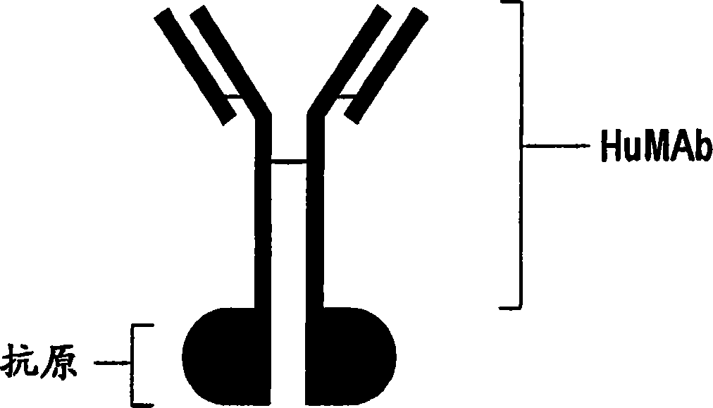
[0246] Design of vaccine conjugates : This construct was generated by linking the βhCG antigen to B11, a fully human antibody that binds to the human macrophage mannose receptor on dendritic cells. Through the method of genetic fusion, the antigen and the heavy chain of the antibody are bonded through covalent bonding, such as image 3 shown.
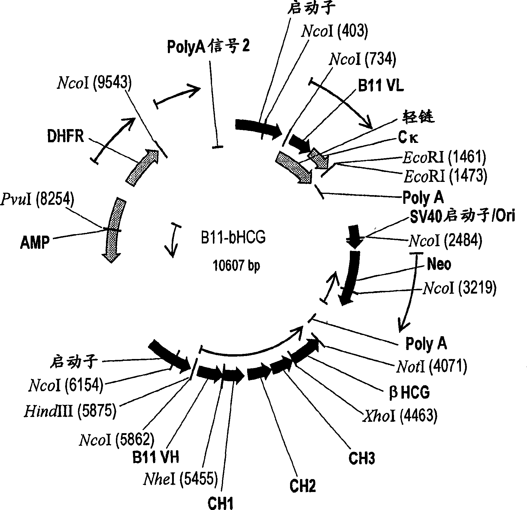
[0247] Recombinant Expression of βhCG-B11 Vaccine Conjugate :Such as figure 2 As shown, a plasmid containing the neomycin gene and the dihydrofolate reductase gene containing the antibody B11 in the heavy chain CH 3 βhCG coding sequence (SEQ ID NO: 9 and 10) fused to the region. The resulting plasmid constructs were transfected into CHO cells using standardized protocols (Qiagen Inc, Valencia, CA). Transfected cells were selected in medium containing the antibiotic G418. Expression is further amplified by growing cells in increasing concentrations of methotrexate. After expansion, ce...
Embodiment 2
[0248] Example 2: Production of B11scfv-βhCG
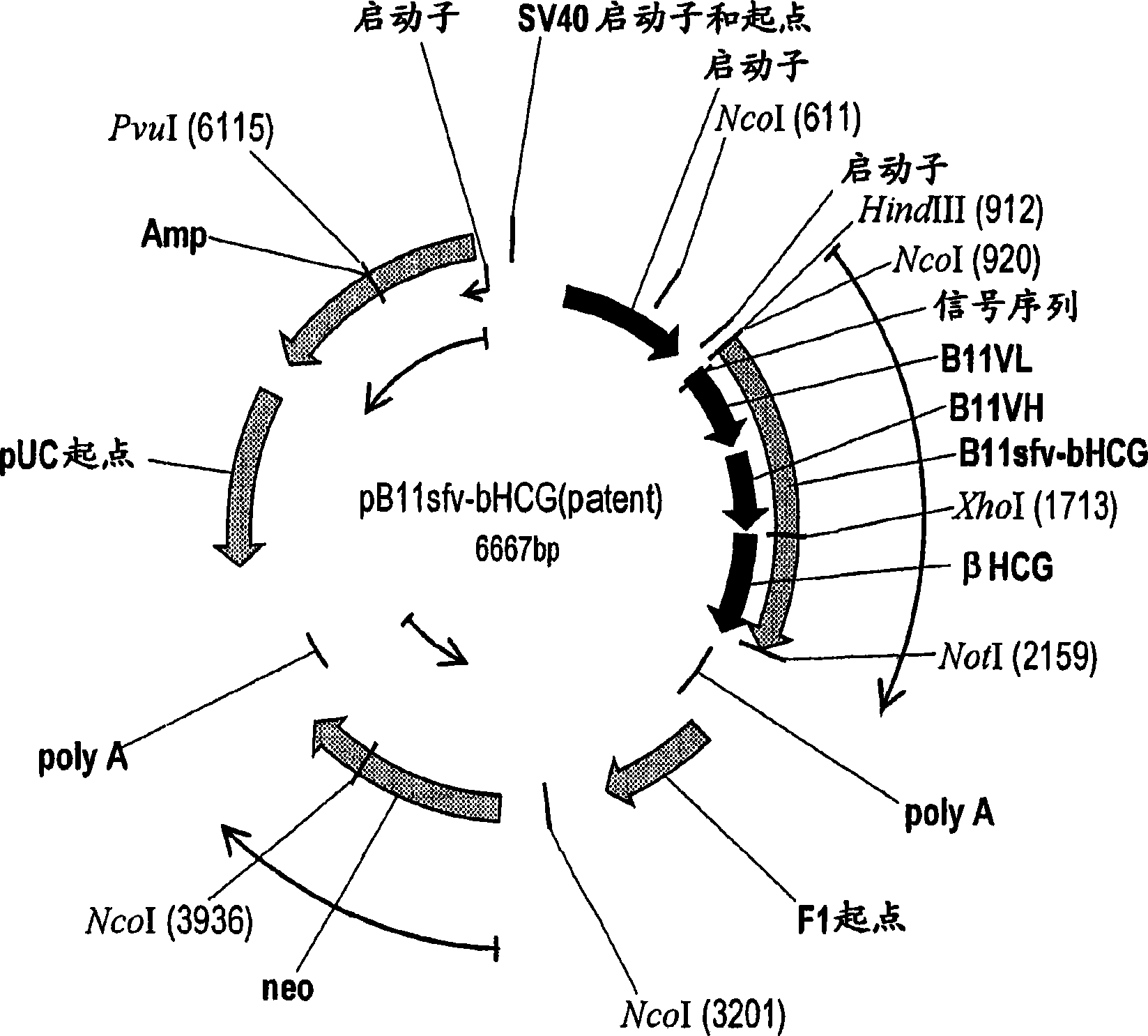
[0249] Design of vaccine conjugates : A second construct was generated by linking the βhCG antigen to a B11 single-chain fusion (ScFv) that binds to the human macrophage mannose receptor on dendritic cells and contains intact human B11 Antibody V L and V H fragments of single-chain antibodies. Through the method of genetic fusion, the antigen is bonded to the carboxyl terminus of B11 ScFv through covalent bonding, such as figure 1 (referred to as B11sfv-βhCG construct).
[0250] Recombinant Expression of B11sfv-βhCG Vaccine Conjugate :Such as figure 1 Plasmids containing the B11sfv-βhCG constructs (SEQ ID NOs: 11 and 12) were generated as indicated. The resulting plasmid constructs were transfected into mammalian cells using standardized protocols (Qiagen Inc, Valencia, CA). Transfected cells were selected in medium containing the antibiotic G418. ELISA was performed to confirm the expression of the B11sfv-βhCG constr...
Embodiment 3
[0251] Example 3: Functional Characteristics of Vaccine Conjugates
[0252] Recognition of antibody-targeted vaccines to their cognate receptors on the surface of APCs is the first step in this drug delivery platform. Flow cytometry studies have demonstrated that the βhCG-B11 and B11sfv-βhCG constructs specifically bind to cultured human DCs expressing MR ( Figure 4 ).
[0253] In situ staining of MR on macrophages in human skin DCs and various human tissue sections was examined using an anti-MR antibody as a probe. Cryosections of human tissue were stained with anti-MR human antibody B11. DCs present in the epidermal layer of the skin were clearly labeled with B11 antibody (data not shown). Binding to DC was noted in the epidermal layer of the skin. Furthermore, immunohistochemical experiments with dendritic cells stained with anti-MR B11 HuMAb in all tissues tested and showed no unexpected cross-reactivity (results not shown). These studies have been repeated with βhCG...
PUM
 Abstract
Description
Claims
Application Information
 - Generate Ideas
- Intellectual Property
- Life Sciences
- Materials
- Tech Scout
- Unparalleled Data Quality
- Higher Quality Content
- 60% Fewer Hallucinations
Browse by: Latest US Patents, China's latest patents, Technical Efficacy Thesaurus, Application Domain, Technology Topic, Popular Technical Reports.
© 2025 PatSnap. All rights reserved.Legal|Privacy policy|Modern Slavery Act Transparency Statement|Sitemap|About US| Contact US: help@patsnap.com


