
Object capture with a basket

a basket and object technology, applied in the field of surgical robotics, can solve the problems of difficult grasping, significant abdominal pain, and reliant on ureteroscopes, and achieve the effect of convenient carrying out a pcnl and easy capture of stones

Active Publication Date: 2018-04-24
AURIS HEALTH INC
View PDF37 Cites 232 Cited by
  • Summary
  • Abstract
  • Description
  • Claims
  • Application Information

AI Technical Summary

Benefits of technology

[0008]This description includes methods and devices for more easily carrying out a ureteroscopy. This description also includes methods and devices for more easily carrying out a PCNL. For ureteroscopy, a basketing device includes a number of independently manipulable pull wires that allow full 360 degree motion of the basket, which makes capture of a stone easier. A central working channel in the basketing apparatus allows a variety of other tools to be placed near a basket to break up a captured stone. Further, a technique for ureteroscopy is described that helps prevent stones from escaping from a basket while the basket is being closed.

Problems solved by technology

Such urinary stones form as a result of concentrated minerals and cause significant abdominal pain once they reach a size sufficient to impede urine flow through the ureter or urethra.
The controls of the ureteroscope are located on a proximal handle of the ureteroscope and accordingly are difficult to grasp as the orientation of the ureteroscope changes.
Accordingly, present ureteroscopy techniques are labor intensive and reliant on ureteroscopes with non-ergonomic designs.
Fluoroscopy increases the cost of the nephrolithotomy procedure due to the cost of the fluoroscope itself as well as the cost of a technician to operate the fluoroscope.
Fluoroscopy also exposes the patient to radiation for a prolonged period of time.
Even with fluoroscopy, accurately making a percutaneous incision to access the kidney stone is difficult and imprecise.
In sum, present nephrolithotomy techniques are costly and problematic for patients.

Method used


Image

  • Object capture with a basket
  • Object capture with a basket
  • Object capture with a basket

Examples


Embodiment Construction

I. Overview

[0041]I.A. Surgical Robotics System

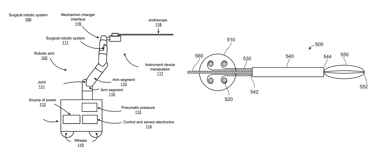
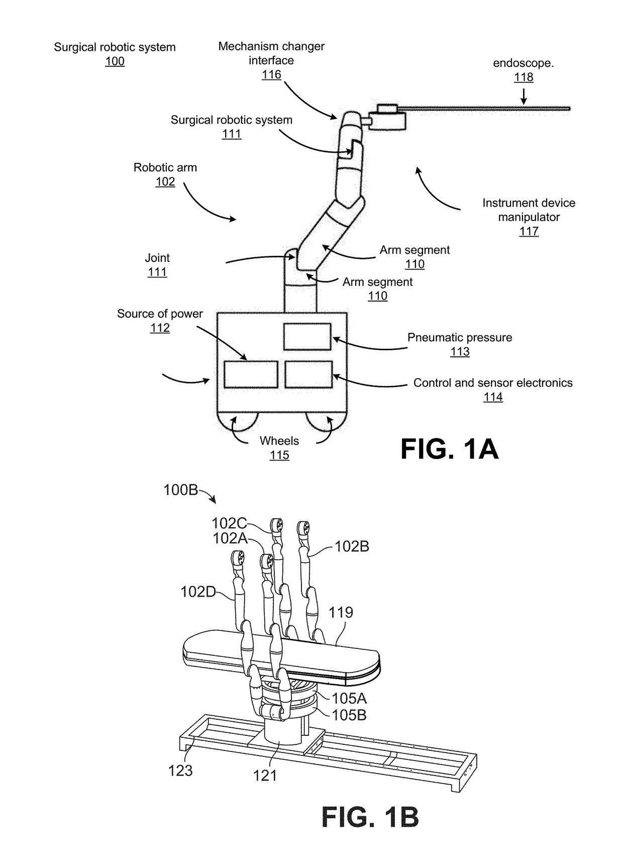
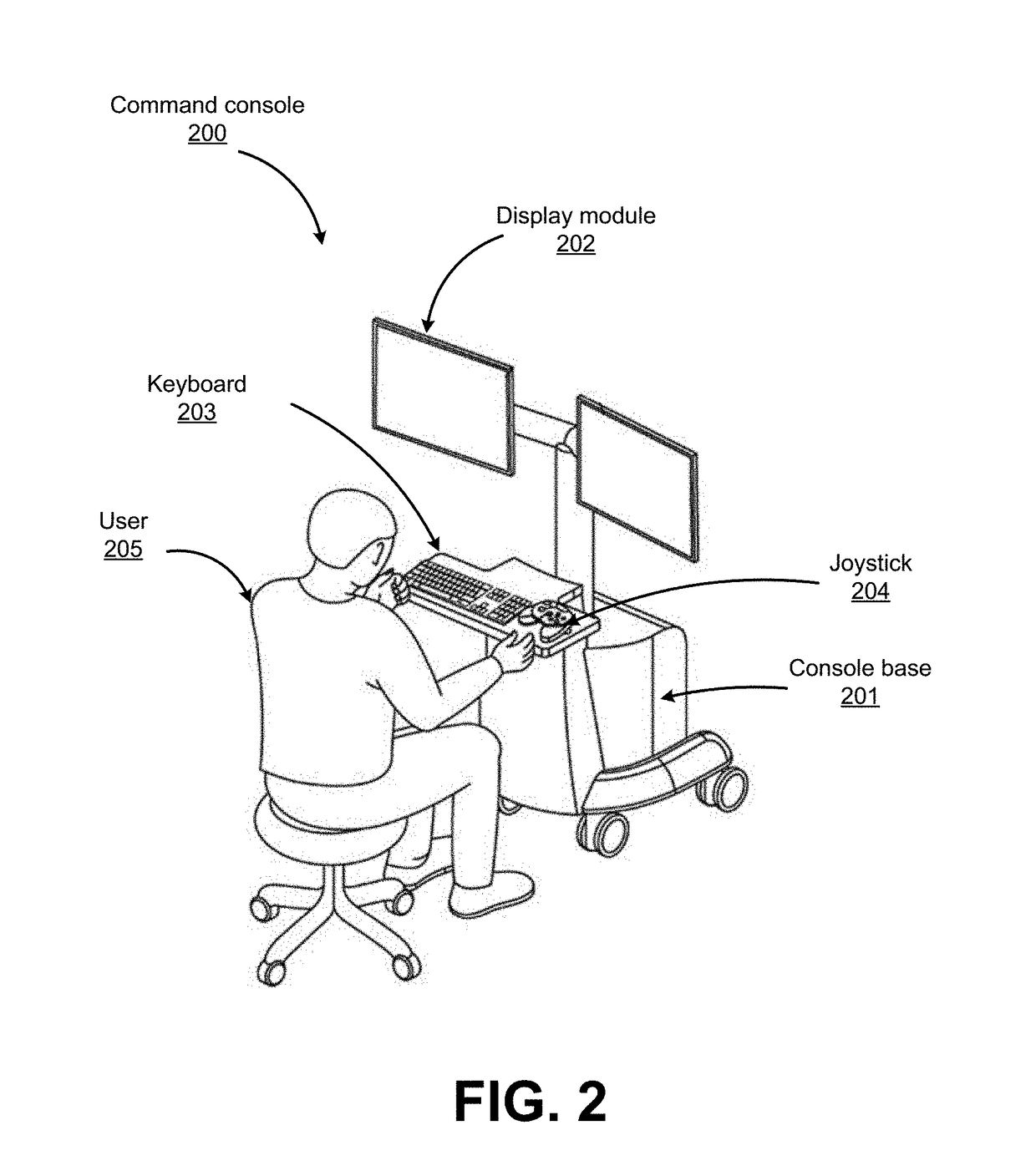
[0042]FIG. 1A shows an example surgical robotic system 100, according to one embodiment. The surgical robotic system 100 includes a base 101 coupled to one or more robotic arms, e.g., robotic arm 102. The base 101 is communicatively coupled to a command console, which is further described with reference to FIG. 2. The base 101 can be positioned such that the robotic arm 102 has access to perform a surgical procedure on a patient, while a user such as a physician may control the surgical robotic system 100 from the comfort of the command console. In some embodiments, the base 101 may be coupled to a surgical operating table or bed for supporting the patient. Though not shown in FIG. 1 for purposes of clarity, the base 101 may include subsystems such as control electronics, pneumatics, power sources, optical sources, and the like. The robotic arm 102 includes multiple arm segments 110 coupled at joints 111, which provides the robotic arm 1...


PUM


Abstract

A method for capturing an object in a cavity in a patient is described. The method includes advancing a ureteroscope into the cavity containing the object. A basket is advanced through a working channel of the ureteroscope. The basket is opened within the cavity and is positioned so as to enclose the object. Then, two actions are performed simultaneously. The basket is collapsed while simultaneously the basket tool is advanced forward so that the object remains within the basket, ideally near the center of the basket, as the basket closes around the object. Once the object is captured, the basket is retracted to remove the object out of the cavity. Further, this process may be automated by having the method carried out by robotic arms acting in tandem, with one or more robotic arms advancing the basket tool or ureteroscope, and another robotic arm collapsing the basket.

Description

CROSS-REFERENCE TO RELATED APPLICATIONS[0001]This application claims the benefit of and priority to U.S. Provisional Application No. 62 / 248,737, filed Oct. 30, 2015, which is incorporated herein by reference.BACKGROUND[0002]1. Field of Art[0003]This description generally relates to surgical robotics, and particularly to lithotomy devices and procedures using a surgical robotics system.[0004]2. Description of the Related Art[0005]Every year, doctors perform thousands of procedures to remove urinary stones from patients' urinary tracts. Urinary stones may include kidney stones found in the kidneys and ureters as well as bladder stones found in the bladder. Such urinary stones form as a result of concentrated minerals and cause significant abdominal pain once they reach a size sufficient to impede urine flow through the ureter or urethra. Such stones may formed from calcium, magnesium, ammonia, ur acid, cysteine, or other compounds.[0006]To remove urinary stones from the bladder and ur...

Claims


Application Information

Patent Timeline
Patent Type & Authority Patents(United States)
IPC IPC(8): A61B17/221A61B1/307A61B1/018A61B17/3203A61B34/00A61B18/26A61B34/30A61B17/3207A61B18/00
CPCA61B17/221A61B1/307A61B17/32037A61B17/320758A61B18/26A61B34/30A61B34/71A61B1/018A61B2217/005A61B2017/2212A61B2017/2215A61B2018/00511A61B2018/00517A61B2034/301A61B2034/302A61B1/00085A61B1/00087A61B1/00149
Inventor NOONAN, DAVID P.URBAN, JR., JOSEPH A.AGRAWAL, VARUN
Owner AURIS HEALTH INC
Learn More