
Visualization and quantification of lung disease utilizing image registration

a technology of image registration and lung disease, applied in the field of visualizing and quantifying lung disease utilizing image registration, can solve the problems of loss of elasticity, high invasiveness, risky and uncomfortable for patients,

Active Publication Date: 2017-09-12
VIDA DIAGNOSTICS
View PDF7 Cites 6 Cited by
  • Summary
  • Abstract
  • Description
  • Claims
  • Application Information

AI Technical Summary

Benefits of technology

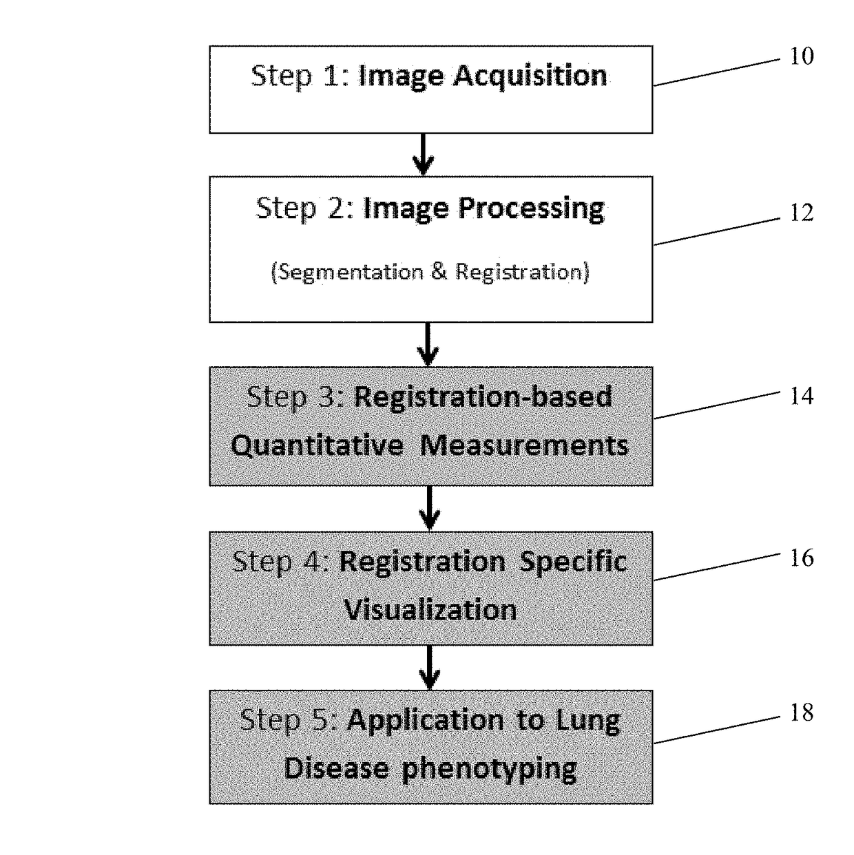
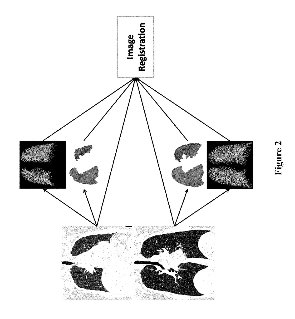
The present invention provides methods and systems for assessing lung function and characterizing lung tissue using volumetric images obtained at inspiration and expiration. The methods involve processing the images to identify known anatomical structures, registering the images to match voxels of the lungs, calculating a continuous probability of lung characteristics at each location in the lungs, and displaying the result as a 3-dimensional image depicting a set of data points. The continuous probability of lung characteristics can be a continuous probability of tissue destruction or a continuous probability of ventilation deficit. The system includes a display, processor, computer-readable medium, and software executable by the processor. The software can be used to classify lung tissue as normal, emphysematous, or having air trapping without emphysema based on the continuous probability of the lung characteristic. The system can also include a graphical user interface for assessing pulmonary function.

Problems solved by technology

Current treatment methods for severe emphysema include lung volume reduction (LVR) surgery, which is highly invasive and can be risky and uncomfortable for the patient.
However, the complex three dimensional structure of the lungs and the diversity of COPD within the lungs of an individual patient makes understanding the disease within each patient difficult.
Emphysema occurs when the lung parenchyma is gradually destroyed, resulting in a loss of elasticity.
Small airway disease, in contrast, is characterized by airway inflammation and remodeling, which can lead to airway obstruction and the trapping of gas within the lung.
However, because of the dynamic nature of the lung due to the expansion that occurs during breathing, assessment of the presence of COPD by this method using one static image is limited.
However, this approach still has limitations.
However, even among these high risk individuals, some individuals may be more likely to develop lung cancer than others.

Method used


Image

  • Visualization and quantification of lung disease utilizing image registration
  • Visualization and quantification of lung disease utilizing image registration
  • Visualization and quantification of lung disease utilizing image registration

Examples


example 1

[0091]Various examples were performed to compare the diagnostic capabilities of the registration methods described herein with other methods. A group of 188 subjects previously diagnosed with COPD at various GOLD stages were used for these examples. The gender, age, weight, and height of the patients in each GOLD stage are shown in FIG. 20. For each subject, full inspiration and full expiration CT images were registered and DPM values were calculated as described above.

[0092]CT based lung function measurements were calculated using the CT images of the subjects, including the EI ratio, the relative volume change between −860 HU and −950 HU (referred to as RVC), gas trapping measurements (referred to as Exp_LAA_856), and percentage of normal tissue computed using a threshold-based registration method (referred to as Galban NormalPerc). Each of these values, as well as the DPM derived normal tissue percentage (DPM_NormalPerc) value, was compared to clinical measurements of lung functi...

example 2

[0101]In another example, the CT images of current and former smokers were analyzed to determine whether the functional assessments described herein could be used to refine existing lung cancer screening guidelines through the use of independent quantitative CT (QCT) biomarkers of lung cancer.

[0102]Full inspiration and expiration CT images were obtained from 322 current or former smokers between the ages of 40 and 89 years old. Of these subjects, 42.5% (n=137) had proven lung cancer, and cancer staging was available for 101 subjects, which included 27.7% stage I (IA=17; IB=11), 18.8% stage II (IIA=11; IIB=8), 27.7% stage III (IIIA=18; IIIB=10), and 25.7% stage IV (IV=26). The images of each patient were processed and registered using nonlinear lung registration, CT functional measurements were calculated, and disease probability measurements were determined as described herein, including DPM_AirTrapPerc, DPM_EmphPerc, and DPM_NormalPerc.

[0103]A logistic regression analysis was perfo...


PUM


Abstract

Methods and systems for assessing lung function using volumetric images obtained at inspiration and expiration. The method may include processing the first and second set of images to identify known anatomical structures of the lungs, registering the first set of images to the second set of images to match voxels of the first set of images to voxels of the second set of images as matched pairs of inspiratory and expiratory voxels, calculating a continuous probability of a lung characteristic at a location of the matched pairs of voxels, and displaying the result on a display. The method may also include classifying lung tissue at each location as normal, having air trapping without emphysema, or being emphysematous.

Description

BACKGROUND OF THE INVENTION[0001]Severe emphysema is a debilitating disease that limits the quality of life of patients and represents an end state of Chronic Obstructive Pulmonary Disease (COPD). It is believed that 3.5 million people in the US have the severe emphysematous form of COPD, and it is increasing in both prevalence and mortality. Current treatment methods for severe emphysema include lung volume reduction (LVR) surgery, which is highly invasive and can be risky and uncomfortable for the patient. New treatment methods for treating emphysema include bronchoscopy guided lung volume reduction devices that aim to close off ventilation to the diseased regions of the lung but maintain ventilation to healthier lung. However, the complex three dimensional structure of the lungs and the diversity of COPD within the lungs of an individual patient makes understanding the disease within each patient difficult.[0002]There are at least two phenotypes of COPD, emphysema and airway obst...

Claims


Application Information

Patent Timeline
Patent Type & Authority Patents(United States)
IPC IPC(8): G06K9/00G06T7/33A61B6/00G06T7/00G06T15/08G06K9/62G06K9/52A61B6/03G06T19/20A61B5/091
CPCG06T7/0012A61B6/032A61B6/50A61B6/5217A61B6/5235G06K9/52G06K9/6201G06K9/6267G06T7/0016G06T7/33G06T15/08G06T19/20A61B5/091G06T2207/10081G06T2207/10088G06T2207/10104G06T2207/30061G06T2219/2012G16H50/30G06F18/22G06F18/24
Inventor YIN, YOUBINGRAFFY, PHILIPPEWOOD, SUSAN A.
Owner VIDA DIAGNOSTICS
Learn More