
Systems and methods for diagnosing strokes

Active Publication Date: 2016-11-08
MG STROKE ANALYTICS INC
View PDF25 Cites 9 Cited by
  • Summary
  • Abstract
  • Description
  • Claims
  • Application Information

AI Technical Summary

Benefits of technology

The invention is about a system and method for diagnosing strokes quickly. This can help doctors provide better information and treatments more quickly for different types of strokes.

Problems solved by technology

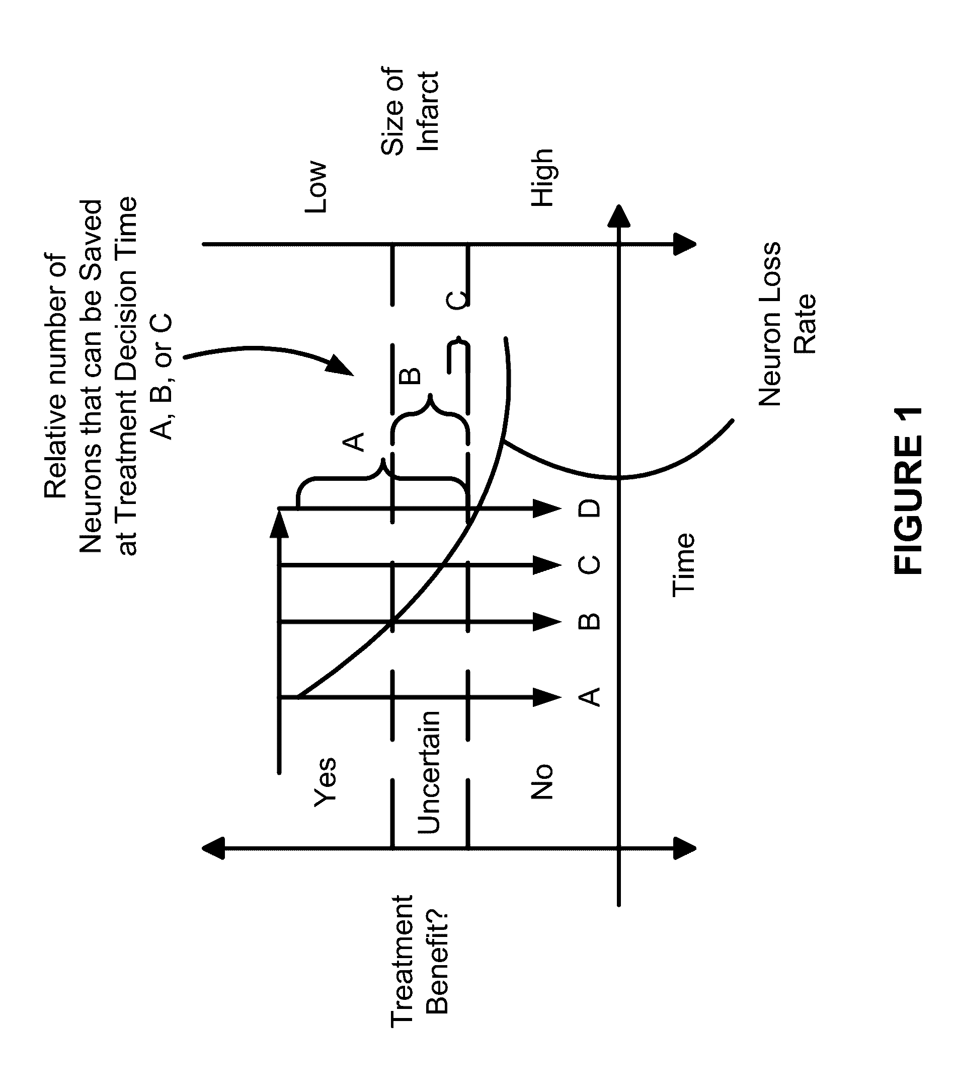
As can be seen, delays in making a decision in the order of only a few minutes can have a significant impact on patient outcome in terms of neural circuitry loss.
CT scans require that the patient hold still during the scan because significant movement of the patient will cause blurred images.
This is sometimes difficult in stroke patients and hence sometimes head restraints are used to help the patient hold still.
That is, as it takes time for contrast agent to flow through the brain tissues and such flow will be very dependent on the ability of vessels to carry the contrast agent, a single snapshot in time does not give the physician enough information to make a diagnostic and / or treatment decision.
It should also be noted that some centers may choose to do a CT perfusion study before the CT angiogram because they feel that the contrast injection from the CT angiogram interferes with the quality of data of the CT perfusion.
The technique is quite vulnerable to patient motion and also requires the patient to hold still for the period.
However, as noted above, there are problems with these procedures.
Lack of consensus in the medical community regarding the interpretation and best practices for treatments based on the CT perfusion maps.i.
In the case of imaging errors, factors including the age and nature of the scanning equipment, set-up and operation (eg. radiation levels) can affect the images and hence their interpretation.
Also, imaging errors can be a result of patient errors include factors such as patient movement, the presence of old infarcts, age of the patient and related factors such atrophy and microangiopathic disease can also affect interpretation.
Image interpretation can be affected by a lack of adequate training including the availability of expertise at a particular facility as well as the pressure physicians may be under during an emergency and the speed with which decisions may be made.
However, at the present time, there is not acceptance that the ASPECTS scores that this software provides are equally or more reliable than physician determined ASPECTS.
That is, there is criticism of this system on the basis that it is not capable of discerning whether the images it is assessing are acceptable images and hence, it will make a determination that does not properly address the quality of the data it is using to make a decision.
As such many such automated algorithms lack precision and / or specificity e.g. depending on the situation especially in a patients presenting very early, an automated software may be able to say that the ASPECTS is somewhere between 4 to 8: this range is such that it does not permit good decision making (following the American Guidelines).
This again would not allow for good and efficient decision making.
Another situation is the patient who at some time went into hypotension / shock before reaching the imaging suite with the result being that the blood supply dropped, the brain died meaning that a poor ASPECTS should be assessed, but prior to subsequent imaging, the blood pressure improved so that by the time the imaging was completed, the collaterals were strong.

Method used


Image

  • Systems and methods for diagnosing strokes
  • Systems and methods for diagnosing strokes
  • Systems and methods for diagnosing strokes

Examples


case 1

A, CTP Procedure

[0128]A 72 year old man presents to the ER at 0820 hours. On examination, he has right hemiplegia and aphasia with an NIHSS of 19. As known, NIHSS is a stroke scale where the NIHSS number is derived from an examination of the patient. The scale range is from zero to 42 with 42 indicating that the patient is dead. Generally, a score of 10 or larger usually means a large stroke.

[0129]A quick examination of the patient is performed (5 min to complete). An IV line is started, blood is withdrawn and the patient is transferred to CT scan. Patient arrives at CT scan at 0840 hours.

[0130]A non-contrast CT scan is performed at 0843 hours. This is immediately seen by the treating physician and it does not show a bleed. The CT technologist immediately sets up for doing a CT angiogram. A CT angiogram is performed (80 cc of contrast is injected). The CTA is completed by 0846 hours.

[0131]The CT technologist gets set up to do a CT perfusion exam (CTP). A localizer is performed and C...

case 2

TA

[0138]A 65 year old presents with slight right sided weakness and slight difficulty in word finding. The NIHSS was 4.

[0139]Patient is taken for a CT, mCTA. The initial non-contrast CT scan is unremarkable. CT angiogram shows an ulcerated plaque at the origin of the left internal carotid artery. No obvious intracranial occlusion is seen. However on the mCTA there is hold up of contrast in one of the branches of middle cerebral artery (MCA) which is detected on the later phases. This allows for detection of an embolus in the M4 branch (one of the distal branches) of the MCA. This has the potential to alter patient management including prognostication, decision on admission as well as whether or not to give thrombolytics.

case 3

A, mCTA, CTP

[0140]A 75 year old woman presents with left hemiplegia at 1520 hours. After assessment the patient is shifted to the CT scan suite. The patient is not cooperative and is not able to hold perfectly still. There is slight amount of motion artifact on the non-contrast CT scan. Some sections have to be repeated. Subsequently, the multiphase CTA is performed. There is again some degree of motion artifact that affects the quality of the scan at the level of the neck and in the second phase. However the intracranial examinations on the first and third phase are of good quality. Subsequently a CTP is performed. However due to patient motion the data is uninterpretable in spite of attempts at motion correction. In this scenario, it is important to note that the uninterpretable data (i.e. marginal data) was not available for consideration until the time the post processing was performed (which as in the example above took approx 30 min beyond the multiphase CTA). The treating tea...


PUM


Abstract

The invention relates to systems and methods for diagnosing strokes. In particular, systems and methods for acquiring timely patient status information are described that enable a physician to make diagnostic and treatment decisions relating to ischemic and hemorrhagic strokes. The systems and methods enable the efficient and quantitative assessment of arterial collaterals within the brain for aiding these decisions in the case of ischemic strokes. In the case of hemorrhagic strokes, the systems and methods are effective in determining if there is a leak and what is the rate of leaking. The systems and methods of the invention can be used to improve the accuracy and confidence of ASPECTS.

Description

FIELD OF THE INVENTION[0001]The invention relates to systems and methods for diagnosing strokes. In particular, systems and methods for acquiring timely patient status information are described that enable a physician to make diagnostic and treatment decisions relating to ischemic and hemorrhagic strokes. The systems and methods enable the efficient and quantitative assessment of arterial collaterals within the brain for aiding these decisions in the case of ischemic strokes. In the case of hemorrhagic strokes, the systems and methods are effective in determining if there is a leak and what is the rate of leaking. The systems and methods of the invention can be used to improve the accuracy and confidence of ASPECTS.BACKGROUND OF THE INVENTION[0002]Ischemic stroke is an acute disease where tissue death (infarction) within the brain of different patients will progress at different rates from the time of the ischemic event. The rate of infarction within a patient depends on a large num...

Claims


Application Information

Patent Timeline
IPC IPC(8): G06K9/00A61B6/00A61B6/03G06T7/00A61B8/00
CPCA61B6/501A61B6/032A61B6/481A61B6/486A61B6/504A61B6/507A61B6/5217G06T7/0016G06T2207/10081G06T2207/20021G06T2207/30016G16H50/30
Inventor GOYAL, MAYANK
Owner MG STROKE ANALYTICS INC
Learn More