
Method for preparing quantitative video-microscopy and associated system

a video-microscopy and video-microscopy technology, applied in the field of image analysis, can solve the problems of limiting the maximum number of markers that may be used in a study, affecting the quality of the sample analysis, and the risk of spectral overlap is higher, so as to reduce subjectivity and inconsistency in sample analysis, and high quality data

Active Publication Date: 2016-03-01
TRIPATH IMAGING INC
View PDF47 Cites 2 Cited by
  • Summary
  • Abstract
  • Description
  • Claims
  • Application Information

AI Technical Summary

Benefits of technology

[0028]Thus, embodiments of the present invention comprise a calibration technique for preparing an imaging system to provide accurate comparisons between multiple images, scenes, fields of view, and / or regions of interest in order to generate high quality data, comprising the necessary information about the sample, while reducing the subjectivity and inconsistency in the sample analysis. Embodiments of the present invention may further provide an apparatus and a computer program product for preparing an imaging system to provide accurate comparisons between multiple images, scenes, fields of view, and / or regions of interest in order to generate high quality data for sample analysis.

Problems solved by technology

However, the maximum number of markers that may be used in a study is restricted by several factors.
For example, the spectral overlapping of the colors used to reveal the respective markers may be a limiting factor because dyes may absorb throughout a large portion of the visible spectrum.
Accordingly, the higher the number of dyes involved in a study, the higher the risk of spectral overlapping.
Further, the spectral resolution of the acquisition device may be a limiting factor and the minimal color shift that the device is able to detect must be considered.
In addition, immunochemistry, as well as chemistry in ISH, are generally considered to exhibit poor sensitivity when quantification of a marker must be achieved.
However, the calibration standard is usually only applicable to a particular specimen, such as a specific cell or a structure of a specific type which is known to exhibit constant features with respect to the standard, and may be of limited utility when applied to a sample of a different nature.
However, such colorimetry studies require extensive sample preparation and procedure control.
Otherwise, the analysis may have to be repeated, starting again from the sample preparation stage, thereby possibly resulting in a costly and time-consuming process.
Accordingly, techniques used in colorimetric analyses of prepared samples are of limited use in the detection and quantification of species of interest due to several factors such as, for example, spectral overlapping, mixing of colors due to spatially overlapping of membrane, cytoplasmic, and nuclear markers, chromatic aberrations in the optical path, limited spectral resolution of the acquisition device, calibration particularities, subjectivity of the detection and quantification process, and inconsistencies between human operators.

Method used


Image

  • Method for preparing quantitative video-microscopy and associated system
  • Method for preparing quantitative video-microscopy and associated system
  • Method for preparing quantitative video-microscopy and associated system

Examples


Embodiment Construction

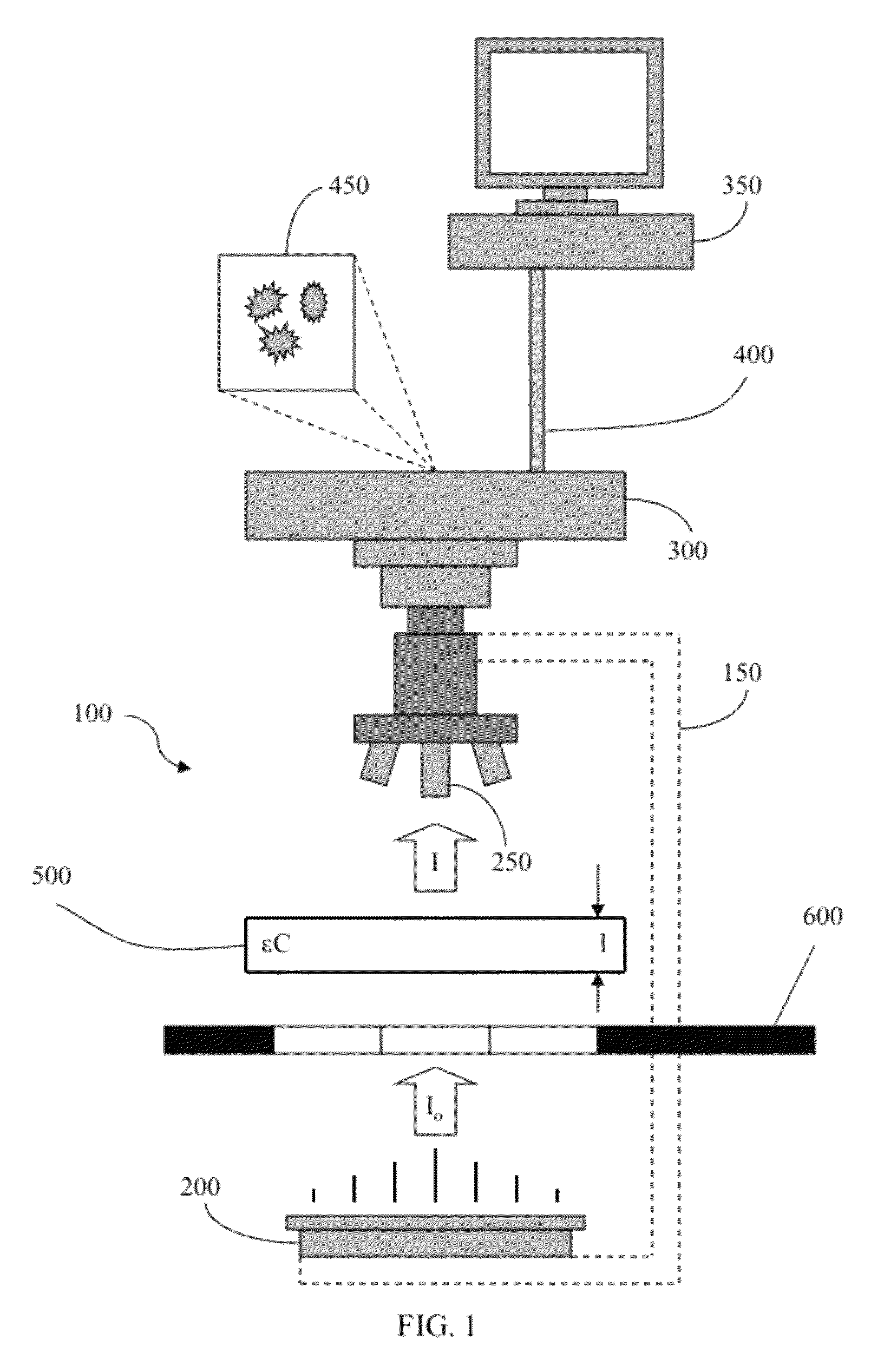
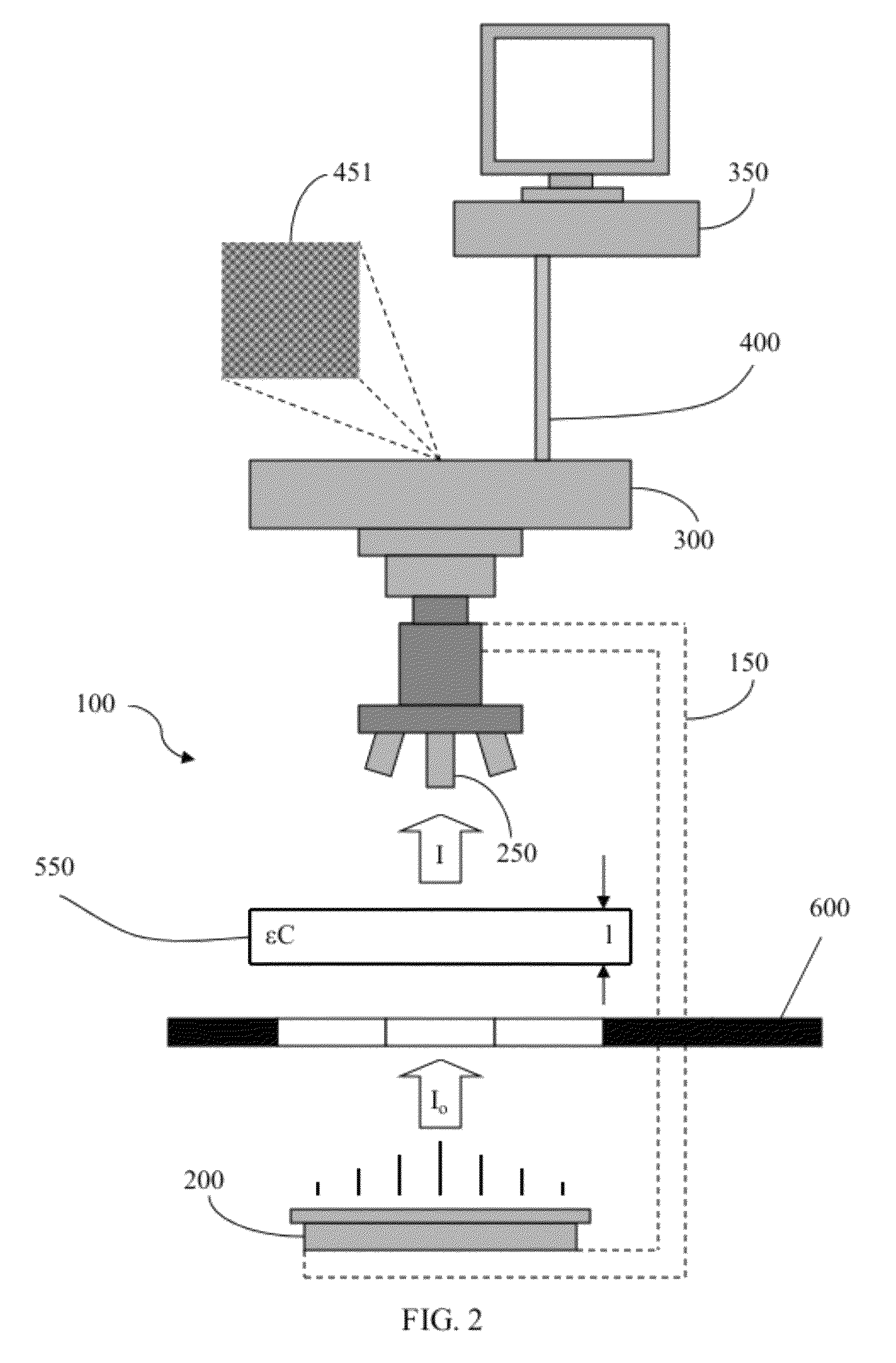
[0016]The above and other needs are met by the present invention which, in one embodiment, provides a method for calibrating an imaging system for analyzing a plurality of molecular species in a sample. The method generally includes acquiring a plurality of images of the sample with an image acquisition device, such as a camera in a video-microscopy system, at a plurality of different wavelengths. The method includes comparing a region of interest associated with at least one of the images acquired at one respective wavelength to a region of interest association with at least one of the images acquired at a different wavelength. Further, the method includes aligning the plurality of images such that the region of interest associated with at least one of the images acquired at one respective wavelength corresponds to the region of interest associated with the at least one of the images acquired at the different wavelength.

[0017]According to one embodiment of the invention, the method...


PUM


Abstract

Embodiments of the present invention are directed to a method for calibrating an imaging system for analyzing a plurality of molecular species in a sample. According to one embodiment, the method comprises acquiring a plurality of images of the sample with an image acquisition device at a plurality of different wavelengths, comparing a region of interest associated with at least one of the images acquired at one respective wavelength to a region of interest associated with at least one of the images acquired at a different wavelength, and aligning the plurality of images such that the region of interest associated with at least one of the images acquired at one respective wavelength corresponds to the region of interest associated with the at least one of the images acquired at a different wavelength.

Description

FIELD OF THE INVENTION[0001]The present invention relates to image analysis and, more particularly, to a method for calibrating or otherwise preparing a video-microscopy system for quantitative video-microscopy in cellular biology and pathology applications and an associated system and a non-transitory computer-readable medium encoded with a computer software program product therefore.BACKGROUND OF THE INVENTION[0002]Effective analysis of microscopic images is essential in cellular biology and pathology, particularly for detection and quantification of genetic materials such as, for example, genes or messenger RNA, or the expression of this genetic information in the form of proteins such as through, for example, gene amplification, gene deletion, gene mutation, messenger RNA molecule quantification, or protein expression analyses. Gene amplification is the presence of too many copies of the same gene in one cell, wherein a cell usually contains two copies, otherwise known as allele...

Claims


Application Information

Patent Timeline
Patent Type & Authority Patents(United States)
IPC IPC(8): H04N17/00H04N7/18G06T5/00G06T7/00
CPCG06T5/006G06T7/0028G06T2207/10056G06T2207/10152G06T2207/20068G06T2207/30024G06T2207/30208G06K9/00134G06T7/33G06V20/693
Inventor MARCELPOIL, RAPHAEL, RODOLPHEORNY, CEDRICK, RENE
Owner TRIPATH IMAGING INC
Learn More