Tissue specimen stage for an optical sectioning microscope

a tissue specimen and microscope technology, applied in the field of tissue specimen stage for optical sectioning microscope, can solve the problems of inconvenient and time-consuming procedure, tissue specimens are generally too thick, and are not useful with optical imaging techniques

Active Publication Date: 2016-01-05
CALIBER IMAGING & DIAGNOSTICS
View PDF84 Cites 8 Cited by
  • Summary
  • Abstract
  • Description
  • Claims
  • Application Information

AI Technical Summary

Benefits of technology

[0009]Accordingly, it is an object of the present invention to provide a window with surface curvature which approximates the shape or curvature of the edges of a tissue specimen when located thereupon so that such edges can contact the window surface, thus avoiding the need to manually position each of the edges.
[0017]The present invention further provides a method for presenting an excised tissue specimen to an objective lens of a microscope comprising the steps of: rotating a window having a surface with curvature upon which an excised tissue specimen is locatable along two rotational axes following of all or part of the curvature of the surface, and supporting the window with respect to an objective lens of a microscope. The rotating step enables the optical geometry to be maintained between the window with respect to the objective lens.

Problems solved by technology

However, since numerous sections must be made from each tissue specimen and then histologically prepared on slides, this procedure is both tedious and time consuming.
This procedure is adequate for obtaining a section which can be placed on a slide for review under a microscope, but is not useful with optical imaging techniques, such as provided by confocal microscopes, which can examine a surgically exposed tissue specimen without the need for traditional microtome sectioning or slide preparation.
One problem with optical sectioning a tissue specimen for Mohs surgery such as by confocal microscope is that the tissue specimen is generally too thick, for example 2-3 mm, to image the edges of the specimen to determine if the specimen contains all of the tumor.
Typically, a confocal microscope is limited to producing adequate images of tissue sections at 100-200 microns.
Although useful, manual positioning needs a skilled technician using a probe to reshape the edges of a thick tissue specimen (e.g., 2-3 mm) to be planar against the planar window surface without puncturing the membrane is a delicate procedure, which if not performed properly can damage the tissue specimen's edges.

Method used


Image

  • Tissue specimen stage for an optical sectioning microscope
  • Tissue specimen stage for an optical sectioning microscope
  • Tissue specimen stage for an optical sectioning microscope

Examples


Embodiment Construction

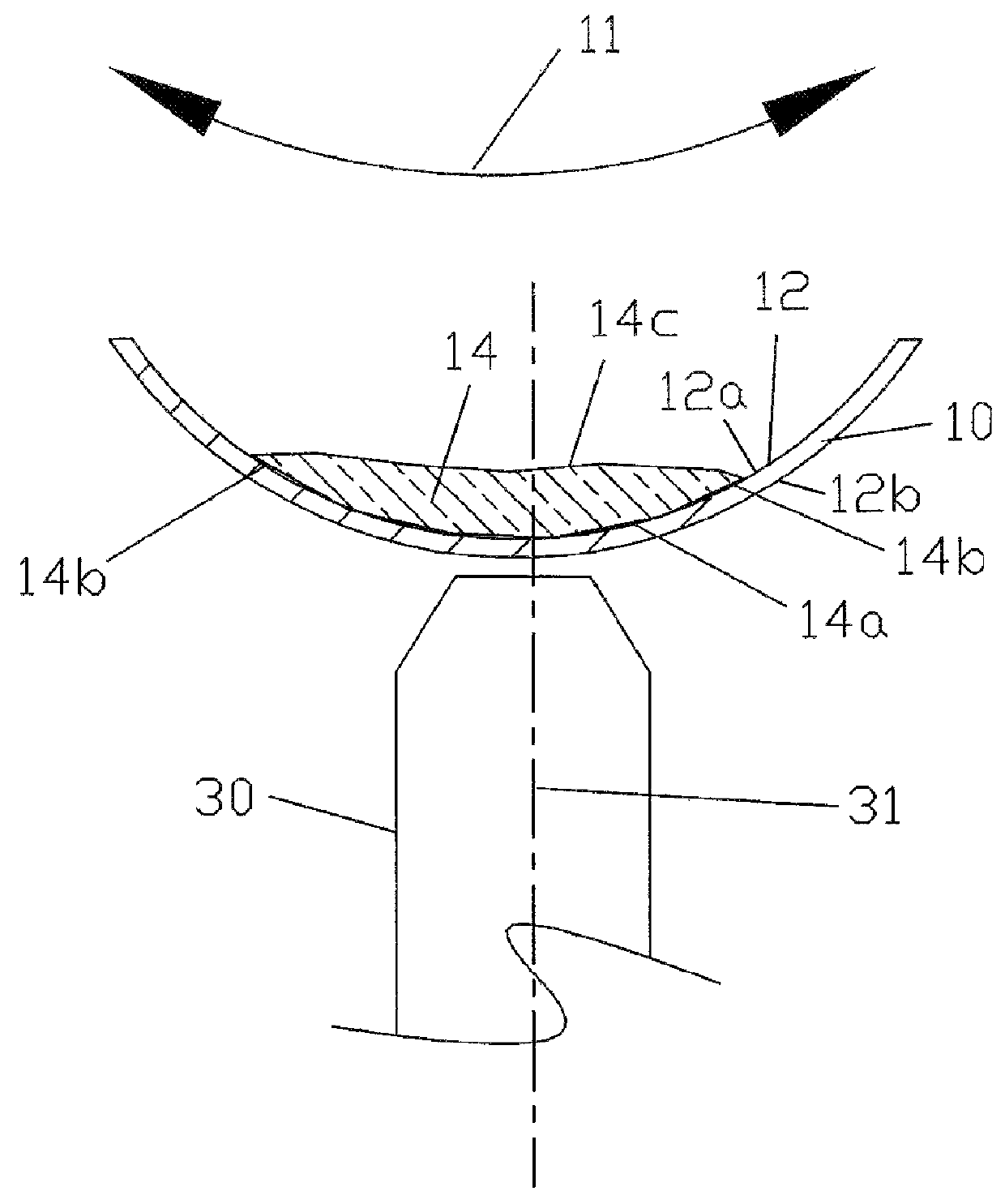
[0031]Referring to FIG. 1A, an example of a window 10 of the present invention is shown having a surface 12 with curvature approximating the shape of the edges 14a and 14b of a tissue specimen 14 when located upon surface 12. Preferably window 10 is rotationally circular symmetric along spherical or concave surface 12. The lower edge 14a and side edges 14b of tissue specimen 14 face surface 12 of window 10, which has a degree of curvature complementary to edges 14a and 14b so that such surfaces can readily contact surface 12 as needed without require individual manual manipulation of side edges 14b. If needed, pressure may be applied onto the top edge 14c of the tissue specimen toward surface 12, such as described below. Tissue specimen 14 and its edges 14a and 14b may be a tissue specimen with edges of interest in removing a tumor, such as in Mohs surgery.

[0032]Although the degree of curvature of the window is complementary to edges 14a and 14b so that such surfaces can contact sur...


PUM


Abstract

A tissue specimen stage is provided having a window with surface curvature upon which an excised tissue specimen is locatable, a carriage having a tissue specimen receptacle to which the window is mounted, and a platform supporting the carriage and presenting the window to the objective lens of an optical sectioning microscope. The carriage is mounted to the platform for movement along two rotational axes so that the carriage's movement follows the curvature of all or part of the window while maintaining the same optical geometry of the window with respect to the objective lens. The window has surface curvature adapted to at least approximate the shape or curvature of the non-histologically prepared tissue specimen to be placed thereupon.

Description

[0001]This application claims priority to U.S. Provisional Application No. 61 / 603,364 filed Feb. 26, 2012, which is incorporated herein by reference.FIELD OF THE INVENTION[0002]The present invention relates to a tissue specimen stage for an optical sectioning microscope, and particularly to a stage (and method) for moving a window with surface curvature adapted to the curvature or shape of the edges and surfaces of a non-histologically prepared tissue specimen. The window is mounted to a container or tissue specimen receptacle that is positioned in a carriage movable along two rotational axes so that different locations along the window are presentable to the objective lens of a optical sectioning microscope for imaging the tissue specimen through the window. Although the present invention is directed for use with a confocal microscope, the stage may be used by other optical sectioning microscopes operable by two-photon or optical coherence tomography as well as microscopes that can...

Claims


Application Information

Patent Timeline
Patent Type & Authority Patents(United States)
IPC IPC(8): G02B21/26G02B21/34
CPCG02B21/26G02B21/34
Inventor FOX, WILLIAM J.DISTASIO, CHRISTOPHER C.
Owner CALIBER IMAGING & DIAGNOSTICS
Why Eureka
Try Eureka