Methods for in vivo identification of cancer initiating cells by multimodality reporter gene imaging
a reporter gene and in vivo technology, applied in the field of in vivo identification of cancer initiating cells by imaging, can solve the problems of challenging in vivo identification of cscs, and achieve the effect of avoiding drug-induced clonal variation and mutation
- Summary
- Abstract
- Description
- Claims
- Application Information
AI Technical Summary
Benefits of technology
Problems solved by technology
Method used
Image

Examples
example 1
Construction of PiggyBac Transduced Multiple Imaging Animal Model
Plasmids and Stable Transfection
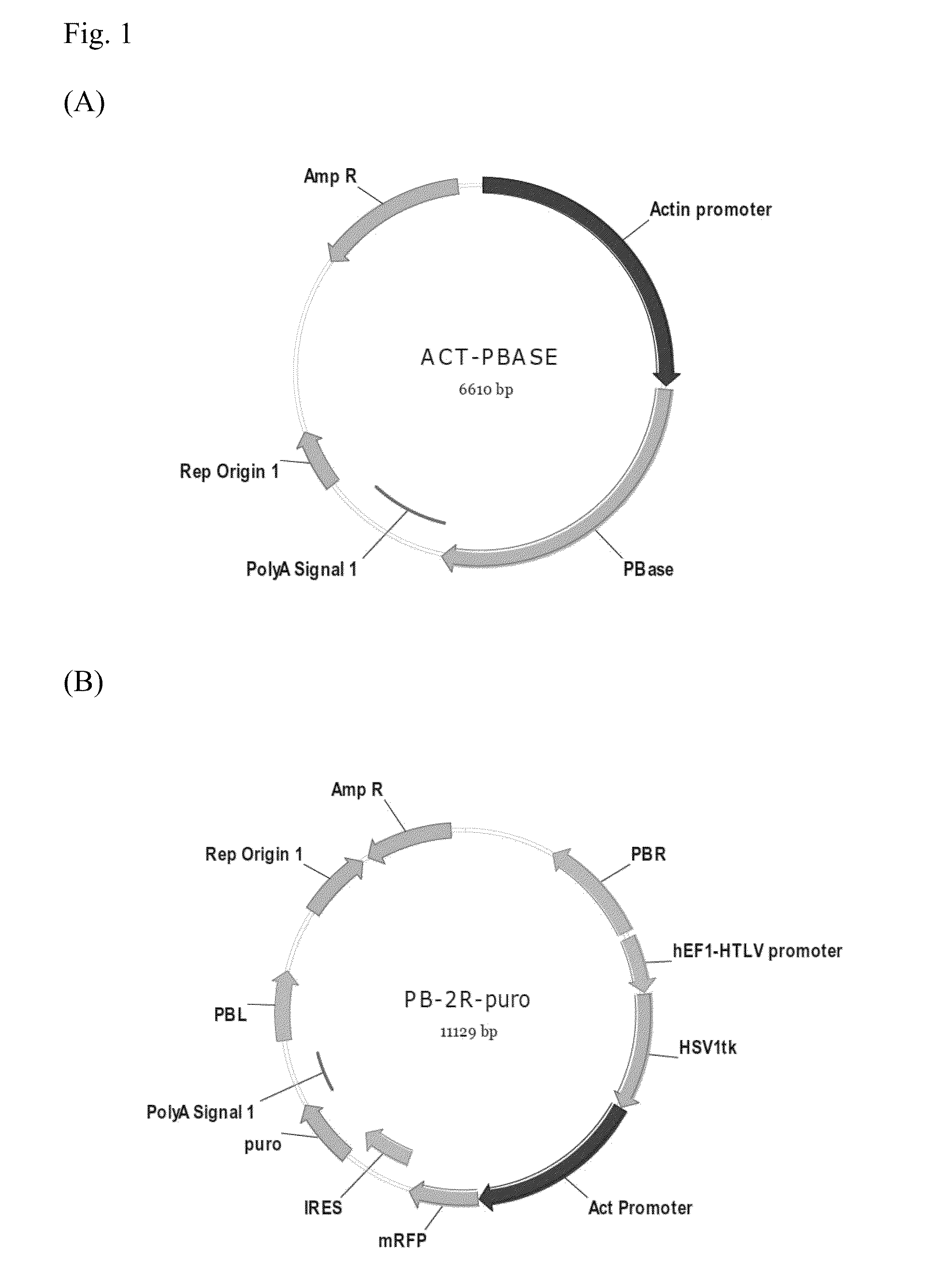
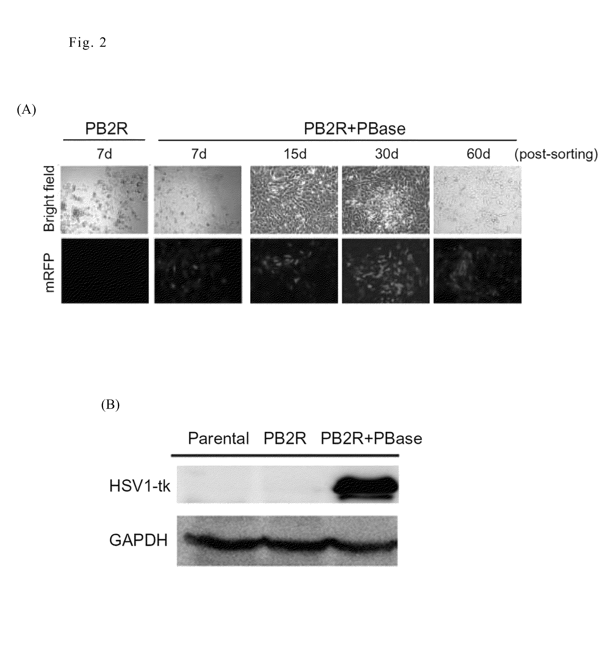
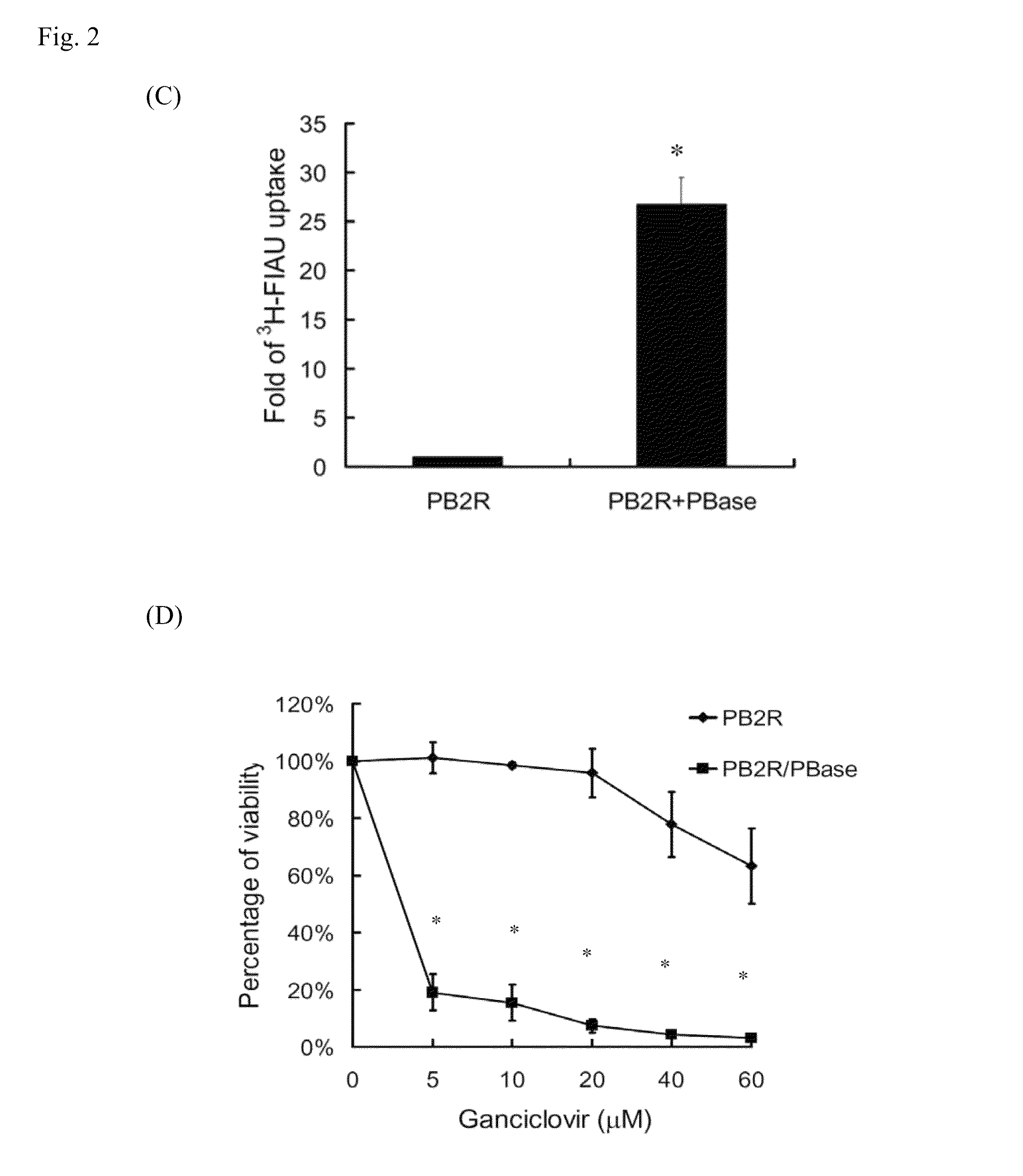
[0020]The PB-tk-mRFP reporter plasmids and Act-PBase helper plasmid were kindly provided by Dr. Congjian Xu (Fudan University, PR China). PB-tk-mRFP was further modified by inserting a puromycin resistance cassette into the BglII and BamHI sites to obtain a new construct named PB-2R-puro. The maps of Act-PBase helper plasmid and PB-2R-puro donor plasmid are showed in FIG. 1(A) and FIG. 1(B), respectively.
[0021]4T1 murine breast carcinomas (a kind gift from Dr. Yueh-Hsing Ou at National Yang-Ming University) were cultured in RPMI1640 medium supplemented with 10% (V / V) fetal bovine serum, 100 U / mL of penicillin, and 100 mg / mL streptomycin. The cell lines were maintained at 37° C. in a humidified incubator containing 5% CO2 and were routinely passaged every two days. For co-transfection, donor plasmid PB-2R-puro was mixed with helper plasmid Act-PBase at the optimal ratio and transfected in...
example 2
Monitor and Identify Solid Tumor Remained Cells by PiggyBac Based Multiple Reporter Gene Animal Model
Syngeneic Tumor Model
[0026]Based on the experimental design, different numbers of 4T1 cells and the derived stable cell lines were implanted into 6-week-old female BALB / c mice (National Taiwan University College of Medicine, Taipei, Taiwan) at subcutaneous positions or in the fat pads. The tumor volumes at subcutaneous positions were measured by caliper every 3 days and calculated using the following formula: Volume=Length (mm)×Width2 (mm2) / 2. An IVIS50 system (Xenogen Inc. Alamda, Calif.) was used to image the expression of mRFP in the 4T1 tumors. The regions of interest (ROIs) were acquired based on the signals emitted from the tumor positions and semi-quantified as photons / sec. Data quantification was analyzed using the IGOR-PRO Living Imaging Software. The animal use protocols have been reviewed and approved by the institutional animal care and use committee (IACUC) of National Y...
example 3
The Remnant Live Cells in Solid Tumors Exhibited the Characteristics of Cancer Initiating Cells
[0033]Cell loss from rapidly growing tumors is caused by intrinsic and extrinsic stresses. However, little is known regarding the properties of the remnant live cells. Because CSCs are resistant to environmental insults, we investigated whether the remnant live cells in solid tumors exhibited the characteristics of CSCs. Based on the results of the multimodality reporter gene imaging described above, ex vivo experiments were performed to isolate the remnant live cells from late-stage tumors formed by 4T1-PB-2R / PBase cells.
[0034]To isolate live cells from the tumors, 4-5 week old tumors were removed and rinsed, minced, and then trypsinized to resuspend the cells. After centrifugation, the cell pellets were cultured in RPMI-1640 medium containing 10% FBS and a high concentration of penicillin / streptomycin solution. The isolated cells were validated by visualization of mRFP expression using a...
PUM
| Property | Measurement | Unit |
|---|---|---|
| V/V | aaaaa | aaaaa |
| V/V | aaaaa | aaaaa |
| V/V | aaaaa | aaaaa |
Abstract
Description
Claims
Application Information
 - R&D
- Intellectual Property
- Life Sciences
- Materials
- Tech Scout
- Unparalleled Data Quality
- Higher Quality Content
- 60% Fewer Hallucinations
Browse by: Latest US Patents, China's latest patents, Technical Efficacy Thesaurus, Application Domain, Technology Topic, Popular Technical Reports.
© 2025 PatSnap. All rights reserved.Legal|Privacy policy|Modern Slavery Act Transparency Statement|Sitemap|About US| Contact US: help@patsnap.com