Fusion proteins for the treatment of allergic diseases
a technology of fusion proteins and allergens, applied in the field of shrimp allergy and fusion proteins, can solve the problems of major and growing medical, social and economic problems, food allergy reactions, etc., and achieve the effect of inhibiting and preventing
- Summary
- Abstract
- Description
- Claims
- Application Information
AI Technical Summary
Benefits of technology
Problems solved by technology
Method used
Image
Examples
example 1
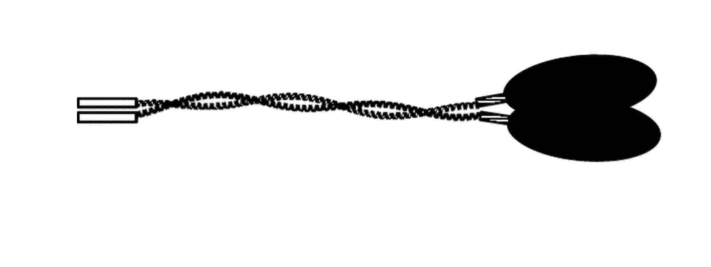
Generation and Characterization of FP; a Fusion Protein Consisting of Shrimp Tropomyosin, a Linker and a C-Terminal FGL2 Peptide CP
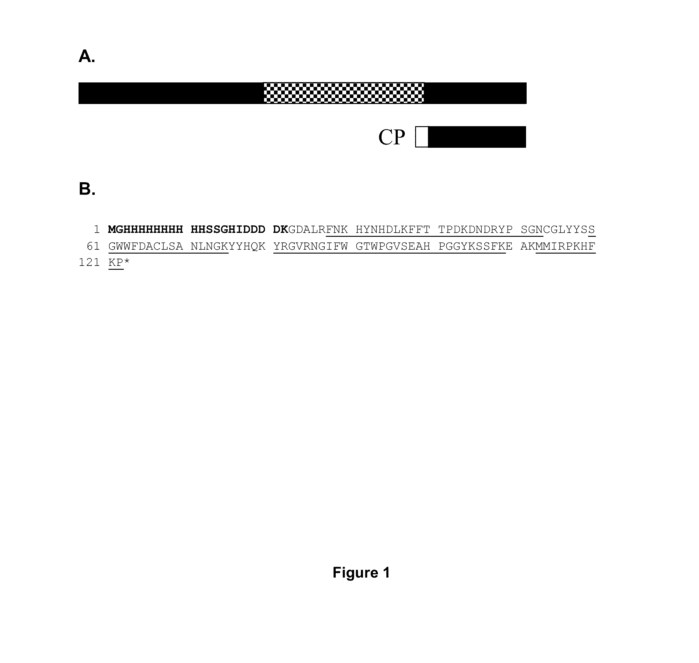
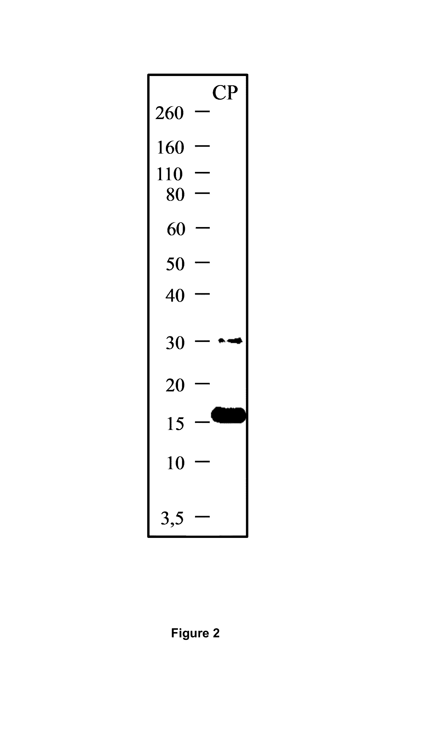
[0218]Design and identification of CP. A C-terminal FGL2 peptide was designed with a FGL2 sequence length of 101 amino acids in order to prevent any possible prothrombinase activity (FIG. 1A). The protein was generated using a E. coli expression system and was purified by IMAC, as described in the methods part. Peptide mass fingerprint analysis of CP provided amino acid recognition of 79% (FIG. 1B). The calculated mass of CP 14.2 kDa. In SDS-PAGE analysis of eluate 1, 2 and 3 after IMAC purification, CP appeared as bands with the approximate weights of 17 kDa (FIG. 2).
[0219]The binding of CP to B-cells was investigated using flow cytometry analysis. FIG. 3 shows that histidin-tagged CP bound on B-cells of normal, non-atopic individuals, while little binding was seen to other cells (data not shown).
[0220]Since CP showed a strong binding to B-cells, this p...
example 2
Generation and Characterization of a Fusion Protein Consisting of a Fragment of Shrimp Tropomyosin and a C-Terminal FGL2 Peptide CP
[0225]Since shrimp tropomyosin has a coiled-coil alpha-helix structure with 5 important IgE binding domains [28], we wanted to investigate whether inclusion of a single IgE binding domain could be of advantage of the structure of the FP. We therefore cloned and expressed five parts of tropomyosin (FIG. 7) and included two of those (region 1 and 5) in a fusion protein with CP. These constructs are hereafter called FP1 and FP5 and thus consist of an N-terminal histidin-tag, the tropomyosin fragment, the short linker, RADAAP (SEQ ID no 12) and CP (FIG. 8). The proteins were expressed in E. coli expression system and results indicated that these proteins also have a dimeric structure (SDS-PAGE, FIG. 9). The shortened fusion proteins were also tested in receptor binding studies (ELISA) and were found to have comparable receptor binding activity as CP and FP (...
example 3
Generation of a Murine Fusion Protein Consisting of Shrimp Tropomyosin and a C-Terminal Mouse FGL2 Peptide mCP
[0227]A murine homologue of human CP (mCP, FIG. 14) was cloned and expressed in E. coli (FIG. 15). CP was included to generate a murine version of FP, and was designated mFP. mFP consisted a N-terminal histidin-tag, shrimp tropomyosin, a short linker and mCP (FIG. 16). SDS-PAGE analysis showed a protein of approximately 50 kDa in size, and presence of proteins of approximately 100 kDa in size, which indicates dimerization of the fusion protein.
[0228]In order to test whether mFP was capable of binding murine B-cells and inhibit allergic responses, an animal experiment was performed. Female inbred C3H / HeJ mice were immunized per-orally with shrimp tropomyosin (Pan b 1) in presence of cholera toxin. This protocol results in tropomyosin sensitized mice with specific serum IgE over time and show anaphylactic reactions after challenge with the specific allergen [29, 30].
[0229]In t...
PUM
| Property | Measurement | Unit |
|---|---|---|
| pH | aaaaa | aaaaa |
| pH | aaaaa | aaaaa |
| pore size | aaaaa | aaaaa |
Abstract
Description
Claims
Application Information
 - R&D
- Intellectual Property
- Life Sciences
- Materials
- Tech Scout
- Unparalleled Data Quality
- Higher Quality Content
- 60% Fewer Hallucinations
Browse by: Latest US Patents, China's latest patents, Technical Efficacy Thesaurus, Application Domain, Technology Topic, Popular Technical Reports.
© 2025 PatSnap. All rights reserved.Legal|Privacy policy|Modern Slavery Act Transparency Statement|Sitemap|About US| Contact US: help@patsnap.com


