
Method for identifying and validating dominant T helper cell epitopes using an HLA-DM-assisted class II binding assay

a technology of helper cell and binding assay, which is applied in the field of biochemistry and immunology medicine, can solve the problems of inability to characterize individual peptides, and inability to detect actual immunogenic peptide sequences

Active Publication Date: 2014-12-23
THE JOHN HOPKINS UNIV SCHOOL OF MEDICINE
View PDF1 Cites 3 Cited by
  • Summary
  • Abstract
  • Description
  • Claims
  • Application Information

AI Technical Summary

Benefits of technology

This approach allows for the efficient identification of physiologically relevant immunogenic helper T cell epitopes, enabling the development of vaccines and immunotherapies by capturing the repertoire of peptides bound to MHC class II molecules, thereby overcoming the limitations of existing methods.

Problems solved by technology

A major problem here is that the actual immunogenic peptide sequences may go undetected because the active region of the immunogenic peptides might not fall within the sequences chosen.
This approach although preferred, is even more tedious, costly, and often unreliable.
Because of the huge number of peptides that need to be tested, and limitations on the responding T cell sources, characterization of individual peptides becomes an impossible task.
However, this approach relies on prediction of peptides that might bind MHC II stably but might not necessarily be immunogenic; hence this method is most unreliable.
Thus, all currently established and commonly practiced methods for identifying immunogenic epitopes are fraught with disadvantages including the fact that they are very tedious, time consuming, costly, and frequently unsuccessful.

Method used


Image

  • Method for identifying and validating dominant T helper cell epitopes using an HLA-DM-assisted class II binding assay
  • Method for identifying and validating dominant T helper cell epitopes using an HLA-DM-assisted class II binding assay
  • Method for identifying and validating dominant T helper cell epitopes using an HLA-DM-assisted class II binding assay

Examples


example i

Materials and Methods

Production of Recombinant Proteins

[0119]Soluble HLA-DR1*0101 and DM were produced as described previously (Stern L I et al., Cell 68:465-77 (1992)) and expressed by “Hi-Five”™ cells (Invitrogen) transduced by recombinant baculovirus containing genes for the α and β chains of human HLA-DR1*0101 and DM lacking their transmembrane and cytosolic domains. The DM α and β chains were genetically modified to possess the FLAG epitope (DYKDDDDK; SEQ ID NO:2) and the cMyc epitope (EQKLISEEDL; SEQ ID NO:3), respectively, at their C-termini. Soluble DM was purified from the culture supernatant by immunoaffinity chromatography with the M2 (anti-FLAG) mAb sepharose resin (Sigma) at pH 6.0, and eluted with FLAG peptide in PBS, further purified by gel filtration chromatography (Superdex 200 HR 10 / 30 column, Amersham-Pharmacia).

[0120]Influenza hemagglutinin HA1 was produced in E. coli. Expression was induced in by the addition of 1 mM IPTG. Bacterial cells were pelleted after 5 h...

example ii

Analysis of Influenza HA Protein

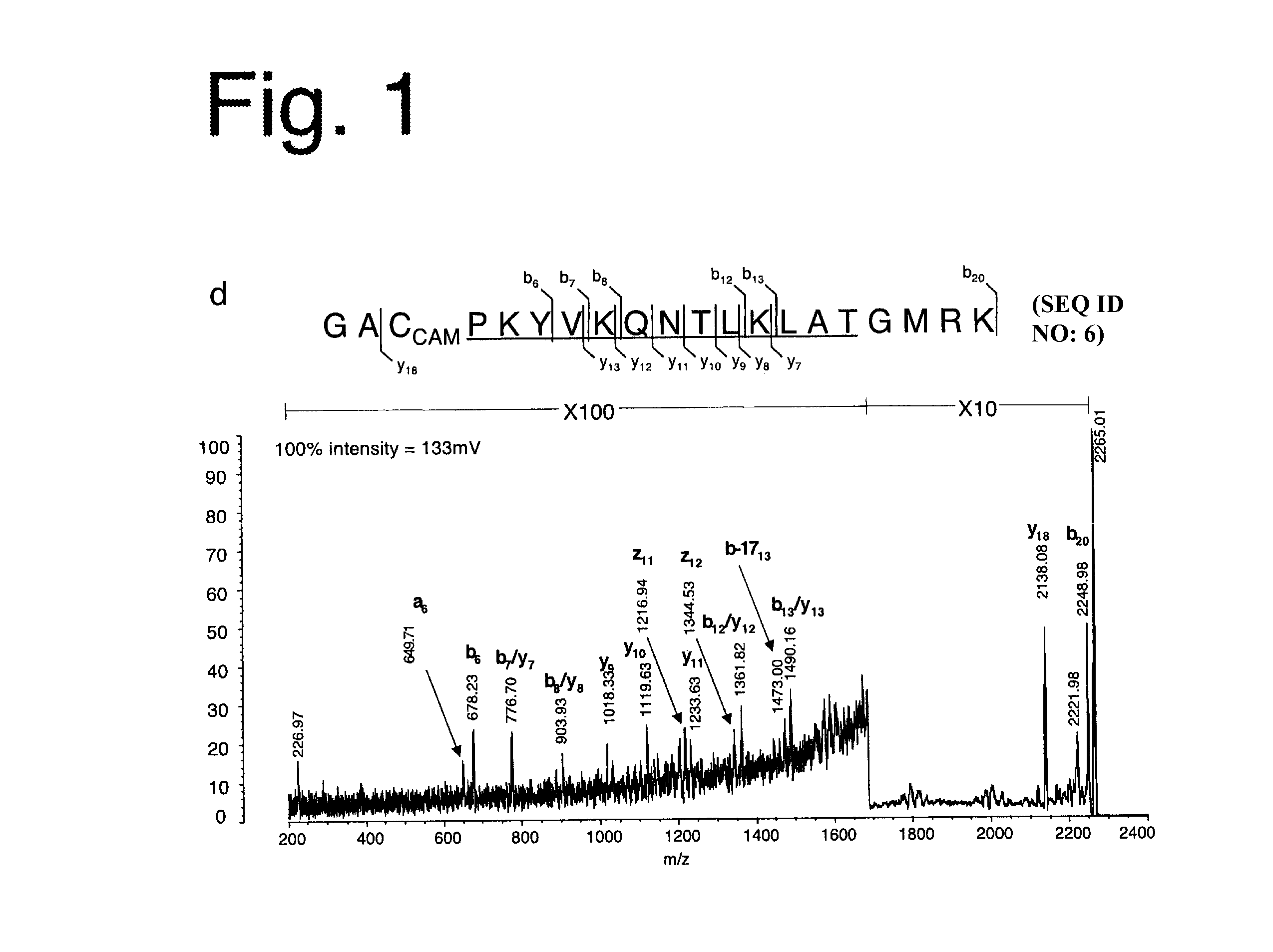
[0124]The influenza HA protein, a well-characterized antigen with a known HLA-DR1-restricted immunodominant epitope, was analyzed. When HLA-DR1+ individuals are infected with influenza strain A / TexasI1177, an epitope made up of residues 306-318 of the HA1 subunit (HA306-318) having a sequence PKYVKQNTLKLAT (SEQ ID NO:5) near its C-terminus emerges as the immunodominant epitope (Lamb, J R et al., Nature 300:66-9 (1982)).

[0125]The present inventors used a recombinant form of HA1 derived from strain A / PR / 8 / 34 to which the A / Texas / 1 / 77-derived HA306-318 epitope was (genetically) fused near the C-terminus where it is naturally present. The resulting protein, susceptible to digestion by cathepsins B and H (FIG. 5), was incubated first with HLA-DR1, with or without DM, and then with cathepsins. HLA-DR1 in the DM-free reaction was induced to adopt a peptide-receptive conformation through pre-incubation with HAy308A, a point mutation of the HA306-318 peptide (...

example iii

Analysis of Type II Collagen

[0130]The present system was next applied to type II collagen (CII), the suspected autoantigen in rheumatoid arthritis (RA) (Trentham, D E et al., J Exp Med 146:857-68 (1977); Yoo, T J et al., J Exp Med 168:777-82 (1988)) The DR1-restricted immunodominant epitope of CII is known to be comprised of residues 282-289, FKGEQPGK (SEQ ID NO:7) (Rosloniec, E F et al., J. Immunol. 160:2573-8 (1998)). Bovine CII was used because it is 98% identical in sequence with human CII and the CII282-289 epitope is conserved.

[0131]Bovine CII (bCII) was digested with matrix metalloproteinase (MMP) 9 (FIG. 6), a neutrophil protease suspected of generating immunologically active CII fragments in RA and known to leave the CII282-289 epitope intact (Van den Steen, P E et al., FASEB J 16:379-89 (2002); Van den Steen, P E et al., Biochemistry 43:10809-16 (2004)).

[0132]The bCII fragments were incubated with HLA-DR1, with or without DM, and were then incubated with or without catheps...


PUM


Abstract

Rational design of immunotherapeutics relies on clear knowledge of the immunodominant epitopes of antigens. Current methods for identifying kinetically stable peptide-MHC complexes are in many cases inadequate for a number of reasons. Disclosed herein is a reductionistic system incorporating known participants of MHC class II antigen processing in solution to generate peptide pools from antigens, including those for which no immunodominant epitope has yet been identified, that are highly enriched for proteolytic fragments containing their immunodominant epitopes. HLA-DM-mediated editing contributes significantly to immunodominance and is exploited in discovering immunodominant epitopes from novel or previously uncharacterized antigens, particularly antigens associated with pathogens, tumors or autoimmune diseases.

Description

RELATED APPLICATIONS[0001]This application is a 371 national stage application of PCT / US2007 / 000180, filed Jan. 8, 2007, which claims priority to U.S. application Ser. No. 60 / 756,882, filed Jan. 6, 2006, U.S. application Ser. No. 60 / 799,281, filed May 10, 2006, and U.S. application Ser. No. 60 / 832,955, filed Jul. 25, 2006, the contents of each of which are incorporated herein by reference.STATEMENT OF RIGHTS TO INVENTIONS MADE UNDER FEDERALLY SPONSORED RESEARCH[0002]This invention was funded in part by grants R01GM53549 and R56AI063764 from the National Institutes of Health, which provides to the United States government certain rights in this invention.BACKGROUND OF THE INVENTION[0003]1. Field of the Invention[0004]The invention in the field of biochemistry and immunology medicine relates to a method of cell-free antigen processing for MHC class II-restricted antigens that exploits the role of HLA-DM in the selection of immunodominant epitopes and permits de novo identification of ...

Claims


Application Information

Patent Timeline
Patent Type & Authority Patents(United States)
IPC IPC(8): C12Q1/37C12Q1/00A61K39/385C07K14/74
CPCC07K14/70539A61P37/04A61K39/145C12N7/00C12N2760/16134C12P21/06
Inventor SADEGH-NASSERI, SCHEHERAZADE
Owner THE JOHN HOPKINS UNIV SCHOOL OF MEDICINE
Learn More