Planning system for intraoperative radiation therapy and method for carrying out said planning

a radiation therapy and planning system technology, applied in radiation therapy, sensors, analog and hybrid computing, etc., can solve the problems of difficult estimation of modifications, difficult to measure the effect of dose measurement, and difficulty in the radiation therapy process

Active Publication Date: 2014-11-04
GMV AEROSPACE & DEFENCE
View PDF18 Cites 5 Cited by
  • Summary
  • Abstract
  • Description
  • Claims
  • Application Information

AI Technical Summary

Benefits of technology

[0025]Generally, the planning system for radiation therapy of the invention comprises at least one central processing unit in which different software packages or modules are executed in order to control and manage all the devices responsible for representing images and touch as well as to gather data relating to the actions performed by the user, simulating an actual surgical procedure; display means for the specialist to view two-dimensional and / or three-dimensional images; a navigator system for locating elements in the three-dimensional space to position the patient and the radiation applicator cone and to adjust it to the pre-planning information; devices for effectively measuring the dose; haptic devices capable of reproducing / simulating the interaction of said radiation applicator with the neighboring tissues in the simulated surgical procedure; and image capture means for documenting the process and checking the tumor tissue remains.
[0029]Based on the resulting image, the system allows the software simulation of inserting the applicator cone in the patient and, in turn, displaying an approximate distribution of the radiation dose received according to the parameters of the applicator cone (diameter, angling, distance to the source of radiation) and of the radiation beam (energy). It further offers the possibility of modifying any of these parameters interactively during said simulation, evaluating different possible scenarios. This operation helps the specialist both in identifying the entry paths towards the tumor and in searching for support surfaces on the tumor bed, further aiding him to have an idea of the homogeneity of the applied dose, of the repercussion on healthy tissues or of the depth of the dose.
[0030]The planning system comprises haptic devices for simulating the handling of the applicator cone, which will further have means for providing force feedback to the user in the event of the virtual applicator colliding with the different parts of the patient's anatomy, thus realistically simulating what will be the subsequent surgical operation and allowing foreseeing a large number of contingencies while at the same time, as is obvious, providing fundamental training to subsequently perform said operation on the patient in an optimal manner.
[0031]On the other hand, as a result of the central processing unit, the system instantaneously presents an estimation of the dose which will change in real time as the position of the applicator cone changes during the simulation. The specialist can combine this dose information with the data from prior radiation therapy sessions received by the patient such that the record book reflecting the radiation received by the patient over a determined time period is updated, which prevents exceeding the total recommended doses for each type of healthy tissue.
[0033]In the intra-planning phase, in the operating room, the system allows, by means of the central processing unit, the radiation applicator cone and the navigator, recording the position of the patient with respect to the accelerator and thus referencing the prior planning data. The system will thus know at all times the position of the applicator and the user can configure the arrangement of the elements of the method, patient and applicator cone as planned. The system allows all the calibration mechanisms necessary so that the error between pre-planning and intra-planning is the lowest possible error. Finally, the user can change planning data according to what is observed during the intra-planning phase.
[0037]Additionally, the system allows verifying the data on the planned dose with respect to the actual dose received by the patient using the devices for effectively measuring the dose, which verification will allow establishing a quality measurement of the pre- and post-surgical procedure.

Problems solved by technology

Currently, although there are a number of dosimetry planning tools typically used for external radiation therapy and brachytherapy, this is not the case for IORT, in which given its special characteristics, said tools do not exist, leaving the calculation of the dose to be applied once the patient has already been subjected to surgery up to the surgeon or radiation oncologist and their experience, i.e., once said specialist has access to the tumor which is to be removed or reduced, which, as is evident, results in difficulties in the radiation therapy process and limits both its quality and the subsequent patient progress follow-up.
The reasons which prevent the existence of IORT planning and dosage tools are the difficulties due to the need for retracting or separating the surrounding organs during the surgical process, and due to the extirpation of the tissues concerned, which facts contribute to modifying the morphology of the patient with respect to that observed in the preoperative image studies.
These modifications are very difficult to estimate beforehand during planning with the information from a preoperative CAT scan.
Furthermore, said problem is even more considerable in the case of IORT, because the radiation is based on the emission of electrons, the dosimetry of which is much more dependent on the exact geometric distribution of the tissues than the radiation based on the emission of photons, which is more common in external radiation therapy.
Therefore, said difficulties translate into the following problems when planning:Before the intervention: it is difficult to estimate the doses that each structure exposed to the radiation will receive.During the intervention: dosimetry estimations adapted to surgical findings cannot be performed, such that the planning can be modified and how possible surgical approach alternatives would affect the dosimetry can be evaluated.After the intervention: since quality control images taken during treatment are not available, as is the case of portal imaging (x-ray images acquired for comparison) in external radiation therapy, that would serve as evidence of the patient's situation during the application of the treatment, the process cannot be correctly evaluated and documented.
This means that currently in IORT, a prior dosimetry estimation of the radiation that will be applied is not performed with suitable reliability (comparable to that which is currently obtained in external radiation therapy) and nor are the obtained results (complete extent of the tumor bed, radiation of healthy tissue, .
The documents existing in the state of the art do not solve these problems because they relate to the external radiation therapy process, as in the case of U.S. Pat. No. 3,987,281, or only to algorithms improving the dose calculation, such as U.S. Pat. No. 4,729,099 or U.S. Pat. No. 6,792,073 for example.
However, none of the described systems is oriented towards intraoperative radiation therapy because they are systems integrated in the external radiation therapy process which do not allow reproducing the anatomical modifications experienced by the patient during the surgical process, or calculating the dosimetry for electron radiation in that particular situation of the patient.

Method used


Image

  • Planning system for intraoperative radiation therapy and method for carrying out said planning
  • Planning system for intraoperative radiation therapy and method for carrying out said planning
  • Planning system for intraoperative radiation therapy and method for carrying out said planning

Examples


Embodiment Construction

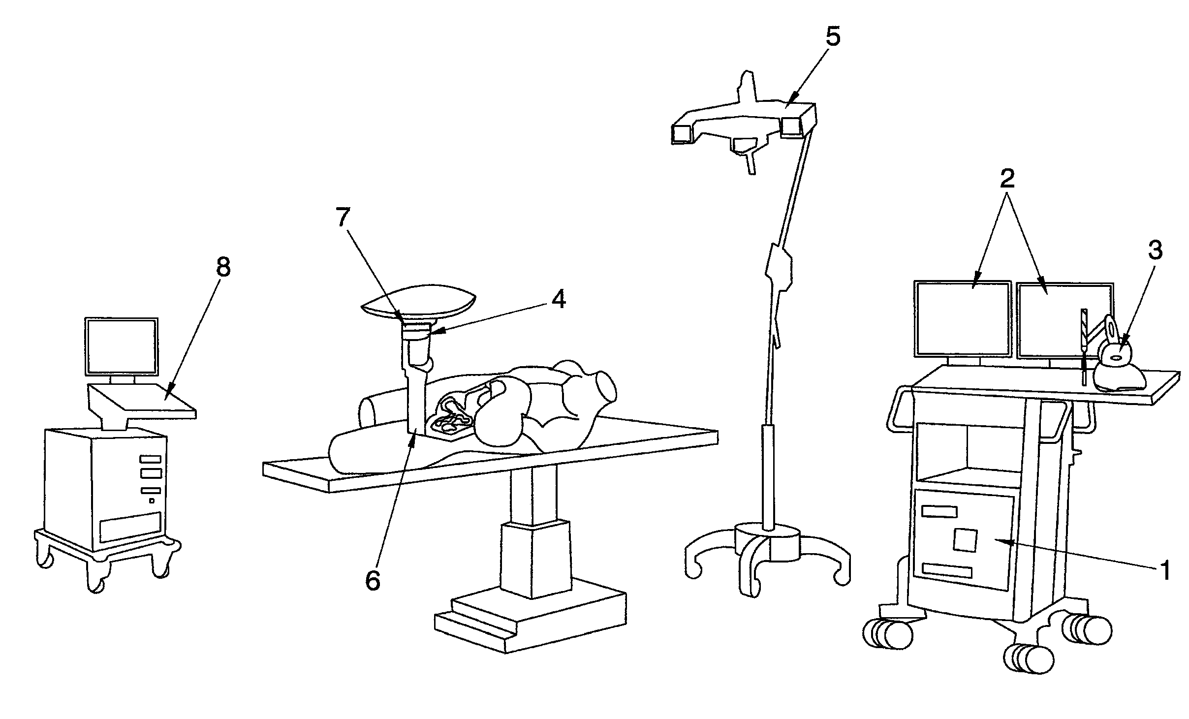
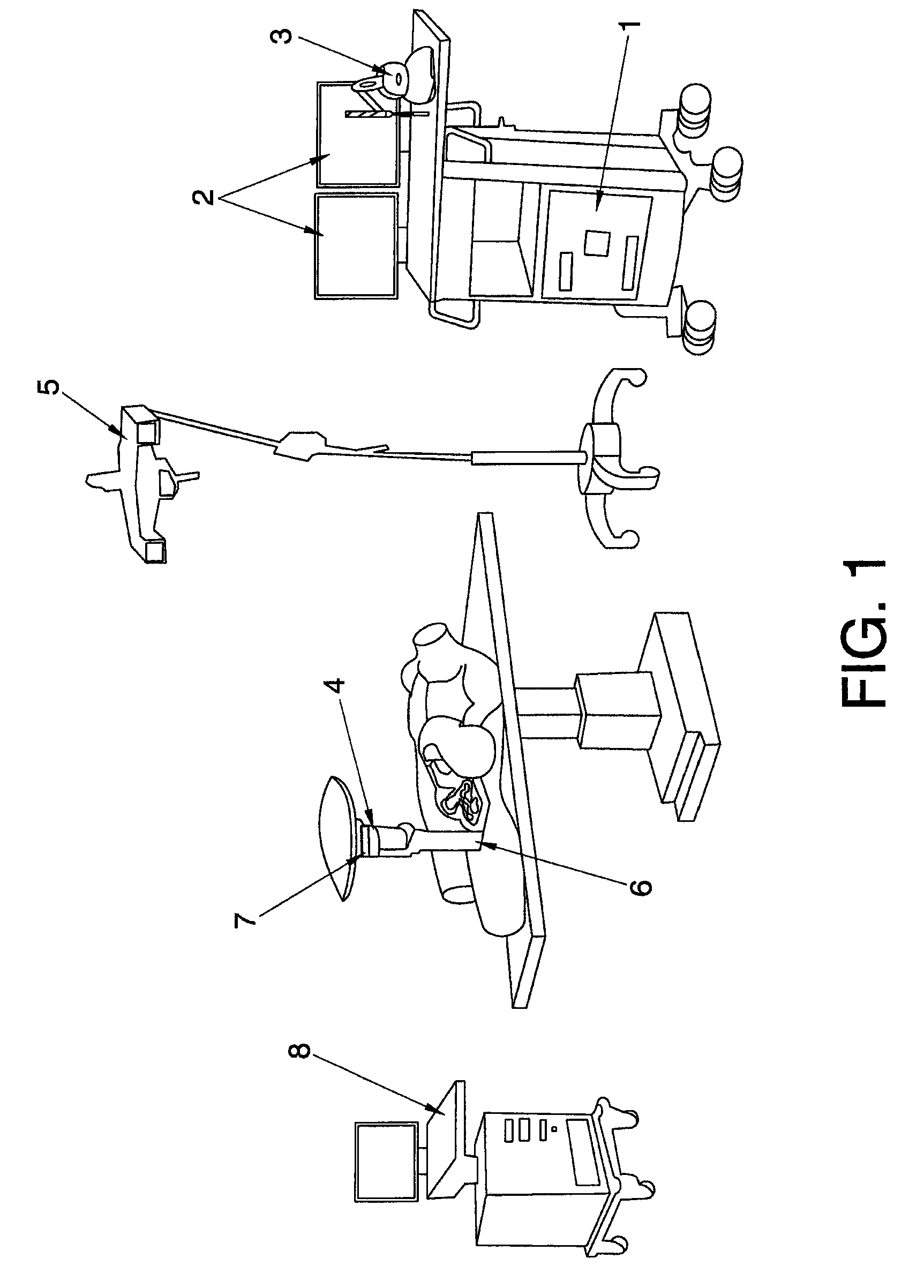
[0042]According to a preferred embodiment described in FIGS. 1 to 3, the planning system for intraoperative radiation therapy of the invention comprises at least one central processing unit or computer (1) for management and control of the rest of the devices, for executing the different software packages or modules responsible for generating the required simulations, for visually representing the patient's anatomy and the doses received, performing the different calculations etc.; devices responsible for representing images such as one or several monitors or screens (2), for example, in which the two-dimensional and / or three-dimensional images which the specialist can observe will be displayed, as well as peripherals responsible for gathering data relating to the actions performed by the user, simulating an actual surgical procedure, such as a computer mouse, a keyboard or haptic devices (3).

[0043]The system of the invention can further comprise a locating system (5) for locating e...


PUM


Abstract

The invention relates to a simulation and planning system for intraoperative radiation therapy and to a method allowing said system to be used for treatment studies, simulation, planning, training and recording, which system generally comprises a central processing unit or computer (1) for management and control and software-based communication with the rest of the devices and the user; one or several monitors or screens (2) for displaying images and peripherals responsible for gathering data relating to the actions performed by said user, a deformation simulation module for the virtual simulation of the deformation produced in the organs and tissues during the process; algorithms for instantly calculating the radiation dose applied during the radiation therapy treatment simulation and means for recording all the activities carried out and generating a full dosimetry report.

Description

TECHNICAL FIELD[0001]The present invention relates to a simulation and planning system for intraoperative radiation therapy and to the process or method allowing said system to be used for treatment studies, simulation, planning, training and recording, and for the suitable dosimetry estimation which will be used in the radiation therapy.[0002]More specifically, the system is designed to measure, calculate, estimate, control, plan and / or simulate the method of radiation that a patient receives during a specific intraoperative radiation therapy treatment.BACKGROUND[0003]Radiation therapy is a form of treatment based on using ionizing radiations (x-rays or radioactivity, including gamma rays, electrons and accelerated particles). Said treatment is commonly used today in oncology treatments for curative or palliative purposes.[0004]Radiation therapy is divided into three types according to the distance of the source of irradiation with respect to the object to be irradiated, these type...

Claims


Application Information

Patent Timeline
Patent Type & Authority Patents(United States)
IPC IPC(8): A61N5/10A61B5/05
CPCA61N5/1038A61N5/1031A61N5/1065A61B19/5244A61N5/1015A61N5/1067A61N5/1071A61N2005/1058A61N2005/1089A61B34/20
Inventor VALDIVIESO CACIQUE, MANLIO FABIOILLANA ALEJANDRO, CARLOS GUILLERMODESCO MENENDEZ, MANUELGONZALEZ-GARZON PASCAU, JAVIERMANUEL CALVO, FELIPEVAQUERO LOPEZ, JUAN JOSE
Owner GMV AEROSPACE & DEFENCE
Why Eureka
Try Eureka