
Coronary artery motion modeling

a motion modeling and coronary artery technology, applied in the field of methods for modeling coronary arteries, can solve the problems of difficult building models of cardiac motion, limited real-time information, and low use rate of 3-d imaging modalities like ct and mri

Active Publication Date: 2014-09-30
SIEMENS HEALTHCARE GMBH
View PDF4 Cites 3 Cited by
  • Summary
  • Abstract
  • Description
  • Claims
  • Application Information

AI Technical Summary

Benefits of technology

The patent describes a system and method for modeling the movement of the coronary arteries in 2-D X-ray images. The system can extract the coronary artery tree for each cardiac phase and maintain the segmentation by tracking the arteries during the cardiac cycle. The system can also map every frame to the specific cardiac phase using ECG values, allowing the shape information of the coronary arteries to be inferred based on their phase. The system has real-time performance and allows the physician to manually adjust the auto-segmented artery tree. The technical effect of this invention is the ability to accurately and efficiently analyze the movement of the coronary arteries in real-time, which can aid in the diagnosis and treatment of cardiovascular disease.

Problems solved by technology

During interventional procedures, 3-D imaging modalities like CT and MRI are not commonly used due to interference with the surgery and radiation exposure concerns.
Therefore, real-time information is usually limited and building models of cardiac motion is challenging.
Coronary motion modeling is an interesting topic and has been researched in depth; however, research on building models from angiographic images is limited.
However, when put into practical use, it appears to be instable and slow.
First, independent artery segmentation can cause a loss of time-continuity in the segmented vessels, which may strip the time-dynamic model of its diagnostic benefits.
For example, a physician may want to modify or mark certain part of the artery segmentation, or even add new vessels that cannot be segmented during the artery tree reconstruction, but the features of an independent artery tree may not be able to be incorporated into the built model.
Second, a segmentation for every phase, including post-processing, can be quite slow and is thus not suitable for real-time applications, like live diagnosis.
Due to issues with existing vessel segmentation algorithms and the lack of contrast in occluded vessels, manual segmentation of certain branches is usually necessary.
Segmenting different cardiac phases independently does not guarantee temporal consistency and is not efficient for occluded branches required manual segmentation.

Method used


Image

  • Coronary artery motion modeling
  • Coronary artery motion modeling
  • Coronary artery motion modeling

Examples


Embodiment Construction

[0030]Exemplary embodiments of the invention as described herein generally include systems and methods for modeling coronary artery motion. Accordingly, while the invention is susceptible to various modifications and alternative forms, specific embodiments thereof are shown by way of example in the drawings and will herein be described in detail. It should be understood, however, that there is no intent to limit the invention to the particular forms disclosed, but on the contrary, the invention is to cover all modifications, equivalents, and alternatives falling within the spirit and scope of the invention.

[0031]As used herein, the term “image” refers to multi-dimensional data composed of discrete image elements (e.g., pixels for 2-dimensional images and voxels for 3-dimensional images). The image may be, for example, a medical image of a subject collected by computer tomography, magnetic resonance imaging, ultrasound, or any other medical imaging system known to one of skill in the...


PUM


Abstract

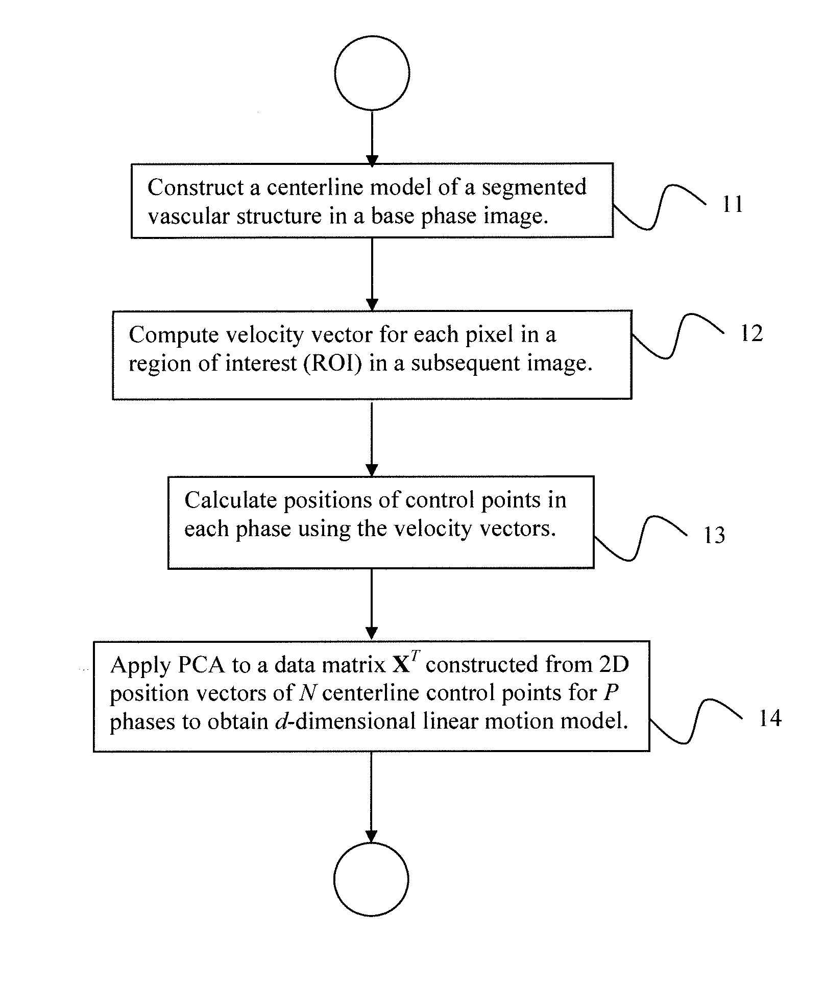
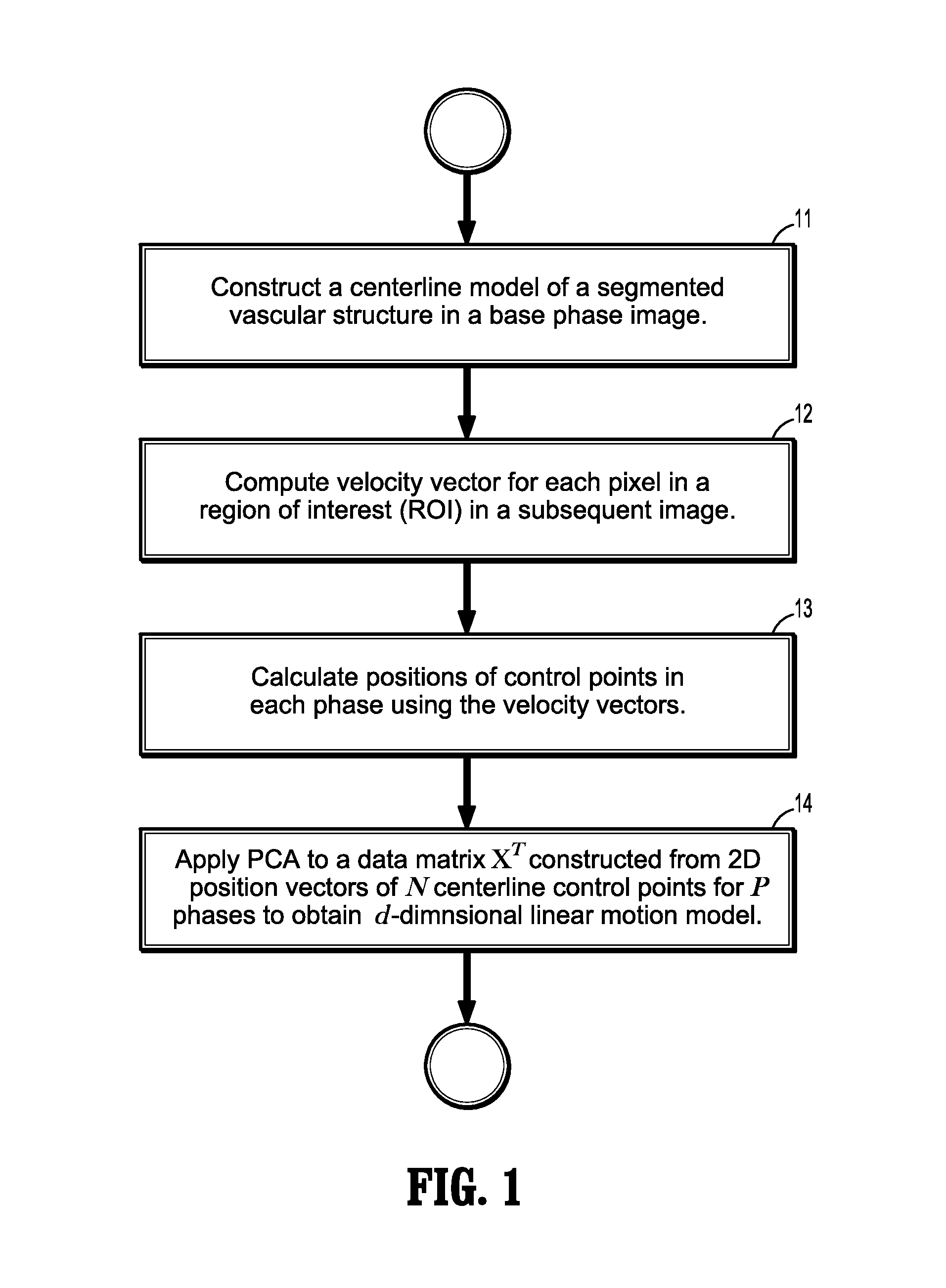
A method for tracking coronary artery motion includes constructing (11) a centerline model of a vascular structure in a base phase image in a sequence of 2D images of coronary arteries acquired over a cardiac phase, computing (12), for each pixel in a region-of-interest in each subsequent image, a velocity vector that represent a change in position between the subsequent image and base phase image, calculating (13) positions of control points in each phase using the velocity vectors, and applying (14) PCA to a P×2N data matrix XT constructed from position vectors (x, y) of N centerline control points for P phases to identify d eigenvectors corresponding to the largest eigenvalues of XXT to obtain a d-dimensional linear motion model {circumflex over (α)}p, in which a centerline model for a new image at phase p+1 is estimated by adding {circumflex over (α)}p to each centerline control point of a previous frame at phase p.

Description

CROSS REFERENCE TO RELATED UNITED STATES APPLICATIONS[0001]This application claims priority from “Coronary Arteries Motion Modeling”, U.S. Provisional Application No. 61 / 540,129 of Sundar, et al., filed Sep. 28, 2011, the contents of which are herein incorporated by reference in their entirety.TECHNICAL FIELD[0002]This disclosure is directed to methods for modeling coronary arteries in digital medical images.DISCUSSION OF THE RELATED ART[0003]Coronary artery disease is a common in many industrialized nations and causes thousands of deaths every year. During interventional procedures, 3-D imaging modalities like CT and MRI are not commonly used due to interference with the surgery and radiation exposure concerns. Therefore, real-time information is usually limited and building models of cardiac motion is challenging. For these reasons, 2-D X-ray angiography is a widely used imaging technique for both diagnosis and for treatment via percutaneous interventions. Quantitative assessment ...

Claims


Application Information

Patent Timeline
Patent Type & Authority Patents(United States)
IPC IPC(8): G06K9/00G06T7/20
CPCG06K9/00362G06T7/2033G06T7/208G06T2207/20076G06T2207/10116G06T2207/10016G06T2207/30101G06T2207/30172G06T7/246G06T7/277G06V40/10
Inventor SUNDAR, HARIGAO, YANG
Owner SIEMENS HEALTHCARE GMBH
Learn More