Method and device for determining oxygen saturation of hemoglobin, for determining hematocrit of blood, and/or for detecting macular degeneration
a technology of hemoglobin and oxygen saturation, which is applied in the field of method and device for determining oxygen saturation of hemoglobin, determining hematocrit of blood, and/or detecting macular degeneration, can solve the problems of reducing the accuracy with which the blood oxygen saturation can be determined, and complicated equations that must be solved to determine the blood oxygen saturation within the retinal vessel, etc., to achieve accurate oxygen saturation determination and increase accuracy
- Summary
- Abstract
- Description
- Claims
- Application Information
AI Technical Summary
Benefits of technology
Problems solved by technology
Method used
Image
Examples
example 1
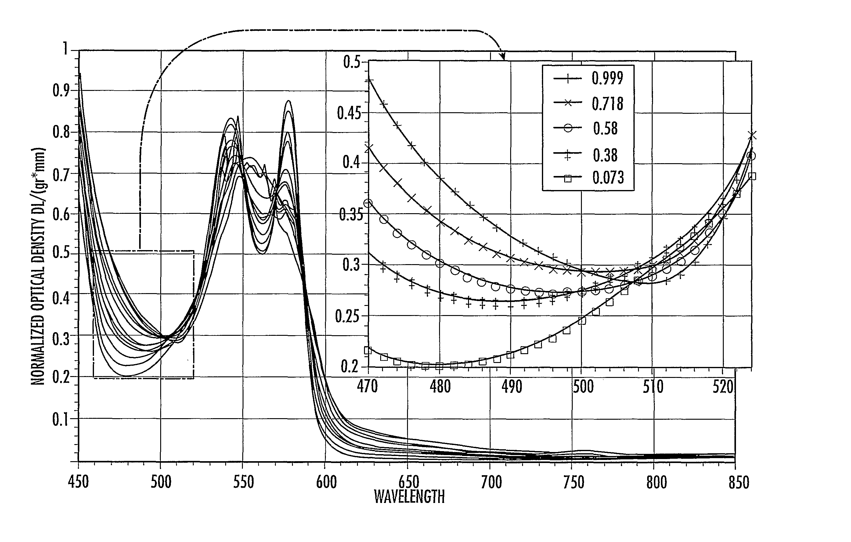
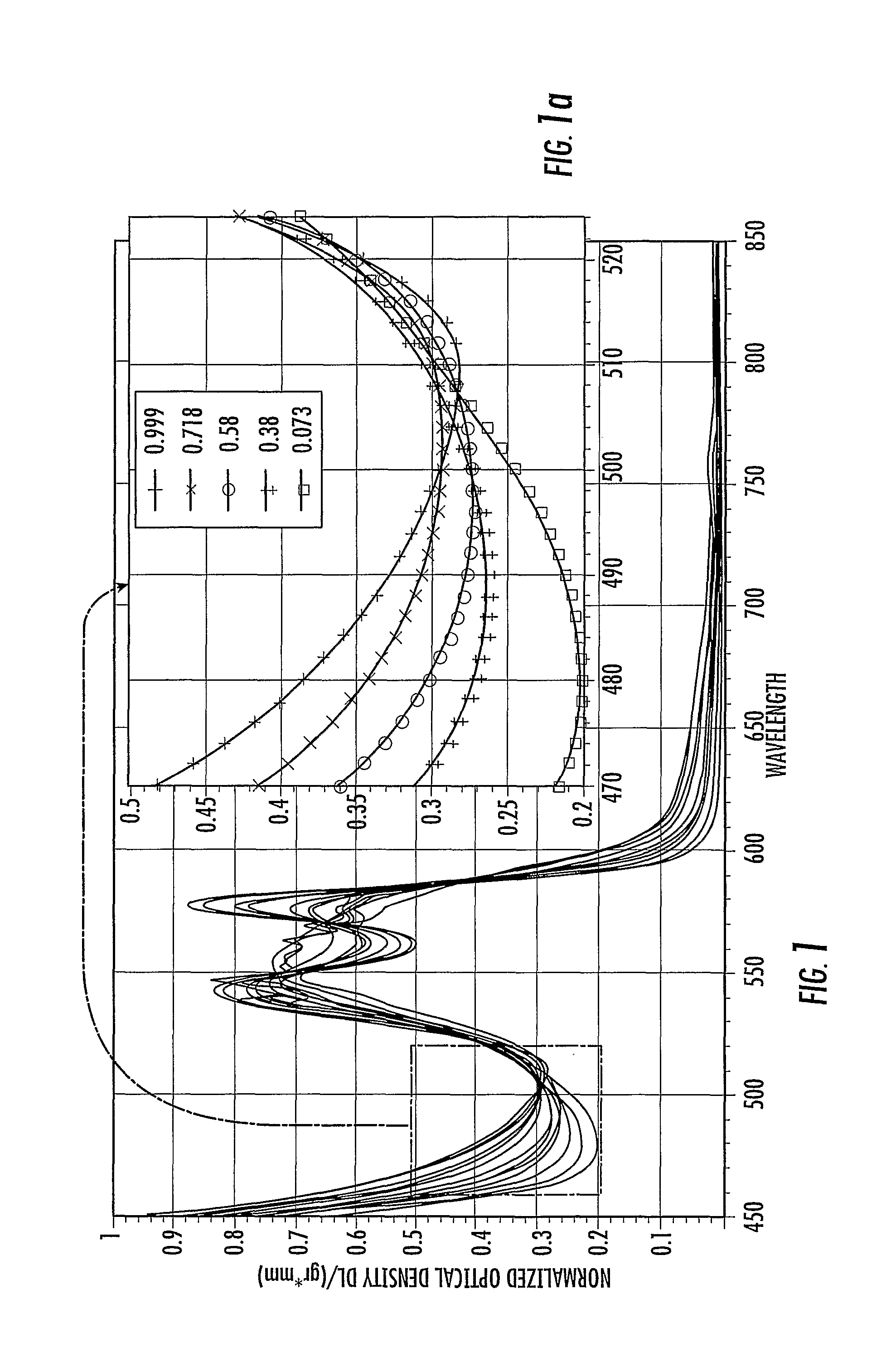
Determination of a Local Minima from Optical Density Measurements with at Least Three Wavelengths
[0040]The parabolic minimum of optical density measure against wavelength may be determined using the following method with three wavelengths:
OD=A*X2+B*X+C (eq. 1)
[0041]wherein OD is the total optical density of the sample; A, B, and C are constants unique to the given spectra; and X is the wavelength of the interrogating light source
[0042]OD is measured at each of three chosen wavelengths within the 460 to 523 nm range and the three constants A, B, and C are determined from the equations resulting from the three wavelengths (three equations, three unknowns).
[0043]Taking the derivative of the above equation (eq. 1) gives the equation:
d(OD) / dx=2AX+B (eq. 2)
[0044]Identifying the minima for this equation where the derivative is zero gives the equation:
0=2AX+B, (eq. 3)
[0045]Solving for X, the wavelength where the optical density in this region is minimal gives the equation:
X=−B / (2A) (eq....
example 2
Detection of a Flux Illuminance Distribution
[0047]This exemplary method allows the quantification of scattered light exiting an eye, which yields a complete measurement of the optical diffused light and the absorptive differences between surface and underlying structure.
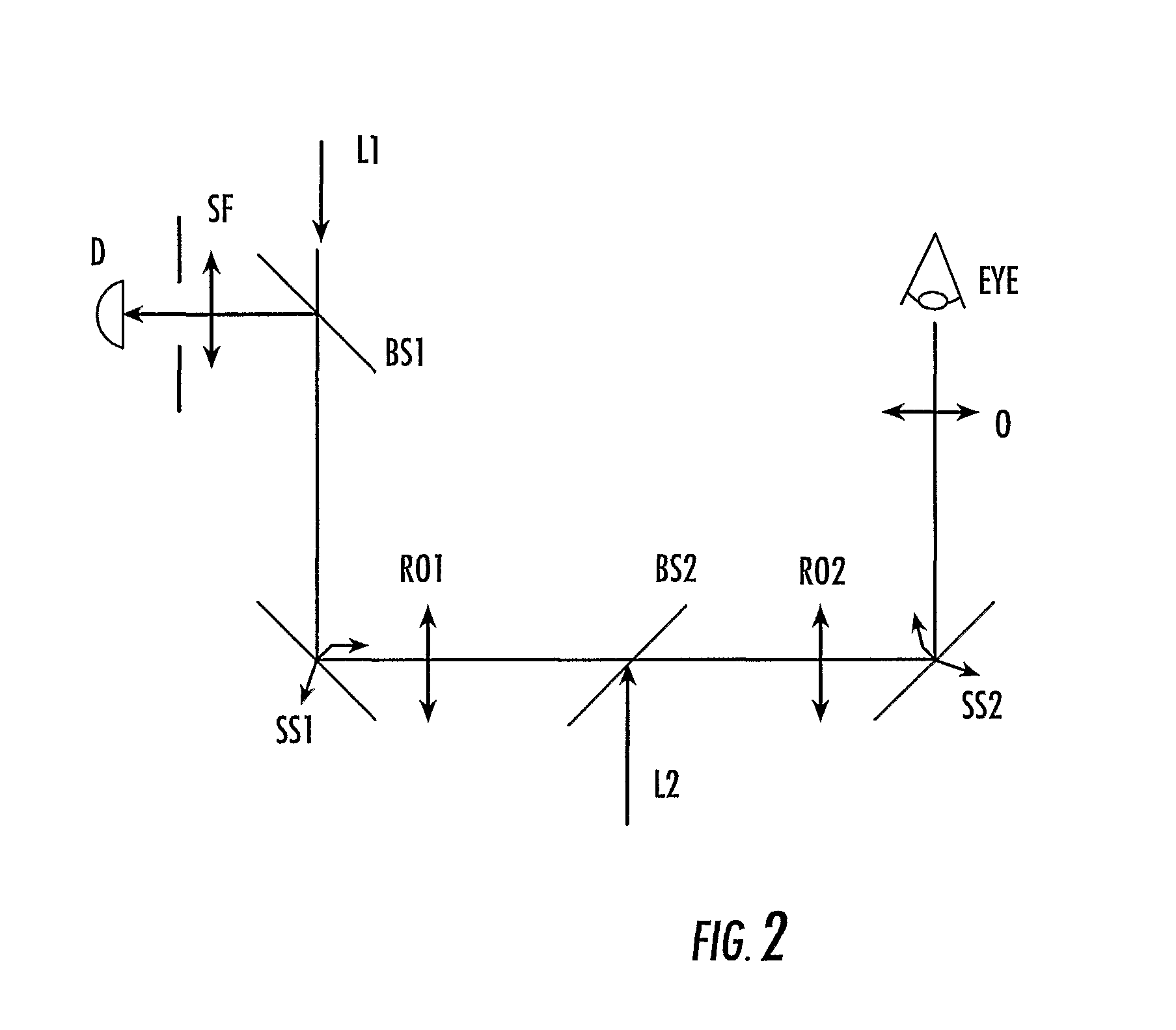
[0048]The main components of the device's optical system (FIG. 2) consist of a (a) tracking laser input path—L1, (b) detector—D, (c) confocal partial filter—SF, (d) beam splitter / combiner for D and L1 paths—BS1, (e) scanning system #1—SS1, (f) scanning system #2—SS2, (g) relay optics for conjugating SS1& SS2—RO (h) probe lasers input path—L2, (i) beam splitter / combiner for D and L2 paths—BS2, and (j) objective lens—O.
[0049]The objective lens, O, collimates the light from either source path L1 or L2 and directs it into the eye. In addition, O also makes conjugate the scanning system SS2 to the pupil of the eye. Motion of the scanning system SS2 scans the probe beam L2 across the back of the retina of the eye. SS2 also...
example 3
Derivation of Improved Method of Oxygen Saturation Measurement
[0050]As noted above, oximetry equations used in the prior art typically assume that there are only two forms of hemoglobin, deoxyhemoglobin and oxyhemoglobin. However, hemoglobin exists as a four molecule grouping that binds to oxygen cooperatively with at least 10 different intermediate combinations of oxygen, hemoglobin and macromolecular structures. In order to more closely model the spectral response of hemoglobin in blood the equilibrium equations for the different oxyhemoglobin intermediates were analyzed. Because hemoglobin has four binding sites that cooperatively bind to oxygen, the resulting equilibrium equations to characterize this relationship were used. Using the equation for oxyhemoglobin saturation S=(((Po23+150 Po2)−1×23,400)+1)−1 the oxyhemoglobin saturation curve was generated. Using the four equilibrium relationships, the equation for saturation S=(0.25C1+0.5C2+0.75C3+C4) / (C0+C1+C2+C3+C4), (C0-C4 are ...
PUM
 Abstract
Description
Claims
Application Information
 - R&D
- Intellectual Property
- Life Sciences
- Materials
- Tech Scout
- Unparalleled Data Quality
- Higher Quality Content
- 60% Fewer Hallucinations
Browse by: Latest US Patents, China's latest patents, Technical Efficacy Thesaurus, Application Domain, Technology Topic, Popular Technical Reports.
© 2025 PatSnap. All rights reserved.Legal|Privacy policy|Modern Slavery Act Transparency Statement|Sitemap|About US| Contact US: help@patsnap.com


