Image capture device for fundus and imaging method

a technology of image capture device and imaging method, which is applied in image analysis, medical science, image enhancement, etc., can solve the problems of reducing the inspection area of the fundus, affecting the examination process, so as to improve the resolution, reduce the cost, and broaden the scope

Active Publication Date: 2014-04-08
ALTEK BIOTECH
View PDF1 Cites 0 Cited by
  • Summary
  • Abstract
  • Description
  • Claims
  • Application Information

AI Technical Summary

Benefits of technology

[0014]Preferably, the image capture device for fundus may further comprise an optical component assembly disposed on the light path between the fundus and the monochrome image sensor, so as to increase or decrease the size of an image of the plurality beams of the reflecting light formed on the monochrome image sensor.
[0020]Preferably, the imaging method may further comprise an optical component assembly disposed on the light path between the fundus and the monochrome image sensor so as to increase or decrease the size of an image of the plurality beams of the reflecting light formed on the monochrome image sensor by the optical component assembly.
[0021]In summation, the image capture device for fundus and the imaging method thereof have one or more of the following advantages:
[0022](1) The fundus image capture device and the imaging method thereof can use the plurality beams of the monochromatic light and base on the principle of different wavelengths having different receiving levels to eyes to project light with appropriate intensity onto a fundus in order to obtain a fundus image with a broader scope effectively.
[0023](2) The fundus image capture device and the imaging method thereof can use the monochrome image sensor in order to lower the cost effectively.
[0024](3) The fundus image capture device and the imaging method thereof can use the monochrome image sensor to improve the resolution higher than the resolution of the color image sensor, under the condition of the same area of the image sensor, or reduce the size of the monochrome image sensor smaller than the size of the color image sensor, under the condition of the same resolution, so as to enhance the image quality or reduce the volume.

Problems solved by technology

Due to a relatively weaker reflection or scattering of the fundus, the imaging process is relatively more difficulty.
Now, the enhanced light source may shrink the pupil, thus reducing the inspecting area of the fundus.
In addition, some prior arts apply a mydriatic agent to the examinee's eye while enhancing the intensity of the light source, but the mydriatic agent usually cause discomfort to the examinee's eye, which is actually troublesome to the examinee.

Method used


Image

  • Image capture device for fundus and imaging method
  • Image capture device for fundus and imaging method
  • Image capture device for fundus and imaging method

Examples


Embodiment Construction

[0032]The technical characteristics of the present invention will become apparent with the detailed description of the preferred embodiments accompanied with the illustration of related drawings as follows. It is noteworthy to point out that the drawings are provided for the purpose of illustrating the present invention, but they are not necessarily drawn according to the actual scale, or are intended for limiting the scope of the invention, and same numerals used in the drawings represent same respective elements respectively.

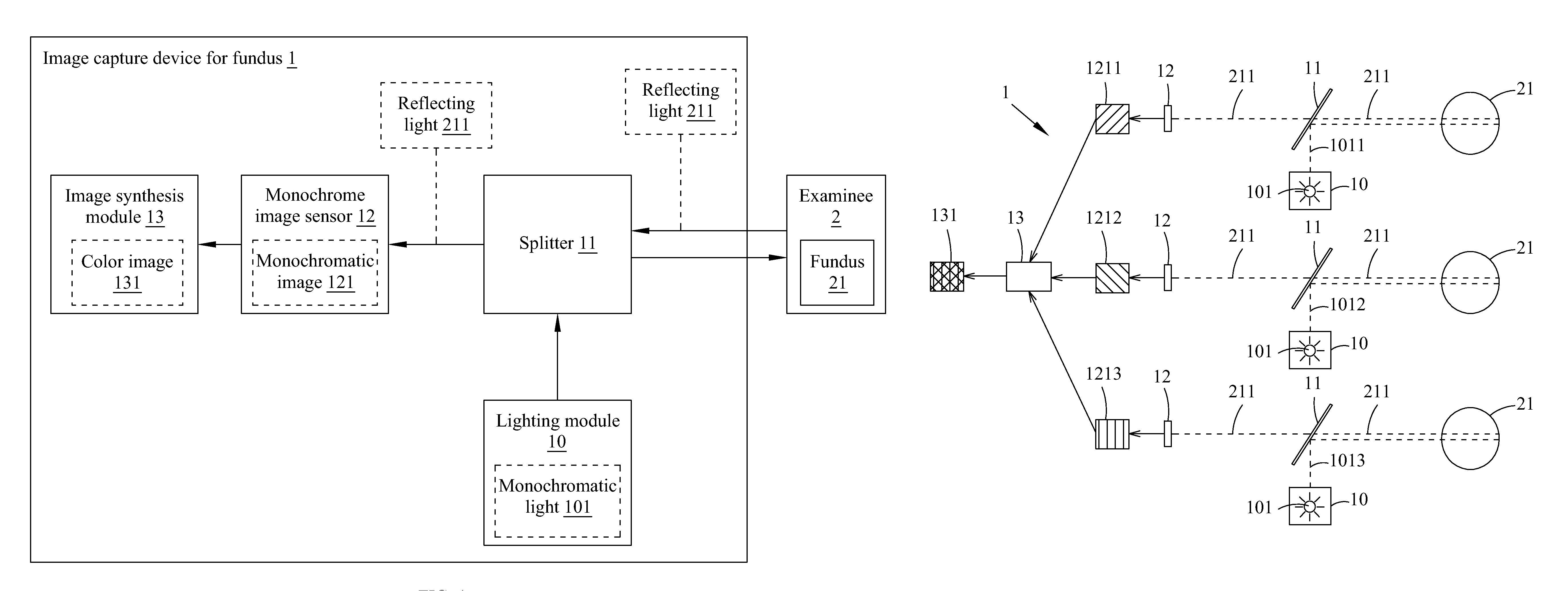
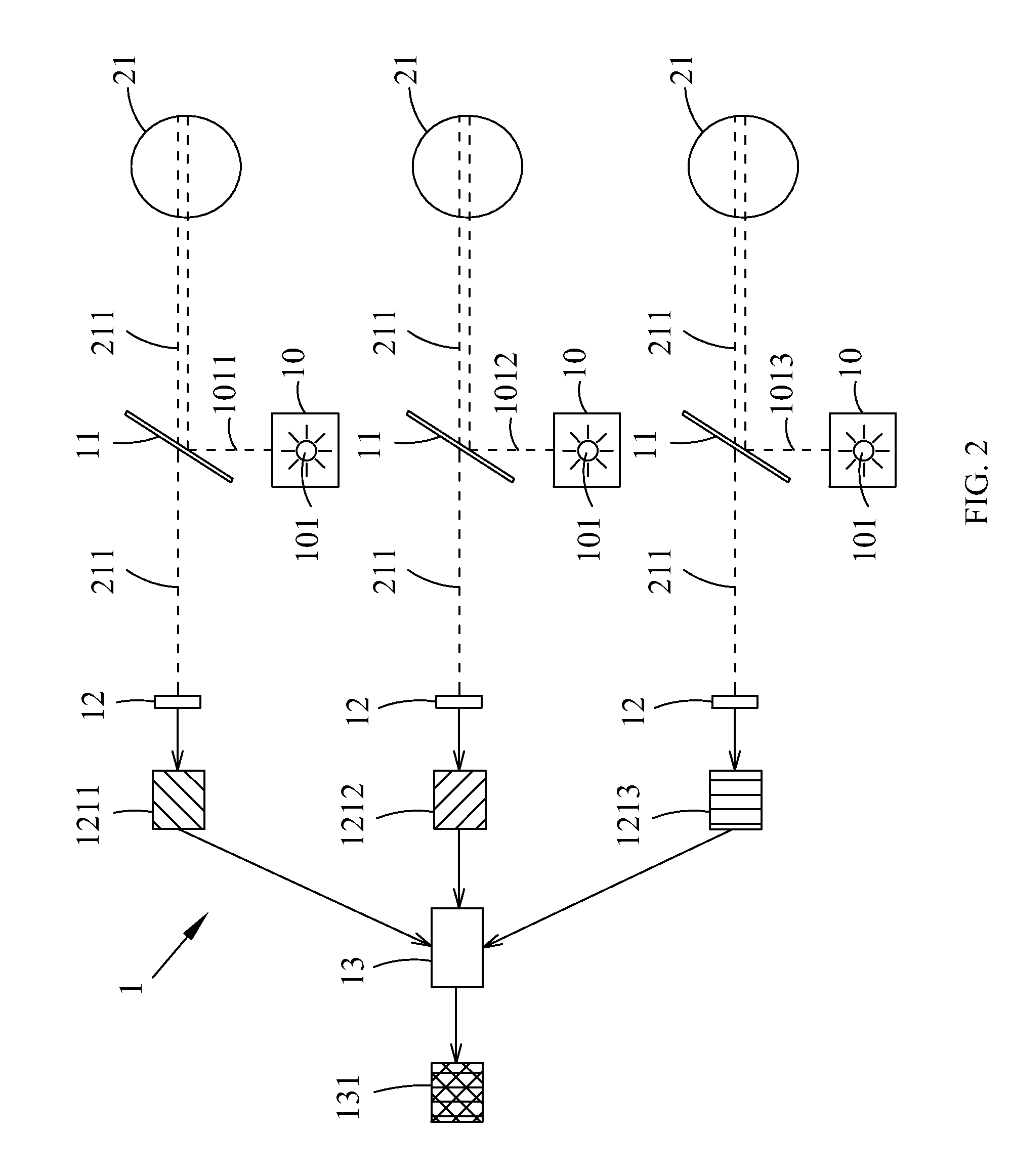
[0033]With reference to FIG. 1 for a block diagram of an image capture device for fundus in accordance with the present invention, the image capture device for fundus 1 comprises a lighting module 10, a splitter 11, a monochrome image sensor 12 and an image synthesis module 13. The lighting module 10 emits plurality beams of monochromatic light 101. The splitter 11 is disposed on a light path of the lighting module 10 and provided for reflecting the plurality ...


PUM


Abstract

This invention discloses an image capture device for fundus and an imaging method thereof. The image capture device includes a lighting module, a splitter, a monochrome image sensor and an image synthesis module. The lighting module emits plurality beams of monochromatic light. The splitter is disposed on a light path of the lighting module. The splitter reflects the plurality beams of the monochromatic light to a fundus of an examinee and forms a plurality of reflecting light respectively through reflection of the fundus. The monochrome image sensor receives the plurality beams of the reflecting light and produces a monochromatic image corresponding to each beam of the monochromatic light. The image synthesis module receives each of the monochromatic images and synthesizes each monochromatic image into a color image. By such arrangements, the invention provides good quality fundus imaging and reduces costs.

Description

CROSS-REFERENCE TO RELATED APPLICATION[0001]This application claims the benefit of Taiwan Patent Application No. 101127723, filed on Aug. 1, 2012, in the Taiwan Intellectual Property Office, the disclosure of which is incorporated herein in its entirety by reference.BACKGROUND OF THE INVENTION[0002]1. Field of the Invention[0003]The present invention relates to an image capture device and an imaging method thereof, in particular to an image capture device for fundus and an imaging method thereof, capable of capturing optical images of eyes for examining the eyes.[0004]2. Description of Related Art[0005]Present eye inspection devices include tonometers, optometry machines and image capture devices for fundus, wherein the image capture device for fundus is an optical inspection device for observing the fundus which is disposed at the rear of a retina of an eye. The image capture device for fundus is mainly used for examining a macular of the fundus and optic nerves to determine whethe...

Claims


Application Information

Patent Timeline
Patent Type & Authority Patents(United States)
IPC IPC(8): A61B3/12A61B3/14
CPCA61B3/14G06T2207/20221A61B3/12
Inventor HSU, CHENG-CHIANG
Owner ALTEK BIOTECH
Why Eureka
Try Eureka