
Method and apparatus for correcting multi-modality imaging data

a multi-modality imaging and data technology, applied in the field of imaging systems, can solve the problems of increasing the dose of x-ray radiation to the patient, image artifacts in attenuation corrected pet images, and increasing the effect of electronic nois

Active Publication Date: 2013-10-08
GENERAL ELECTRIC CO
View PDF26 Cites 7 Cited by
  • Summary
  • Abstract
  • Description
  • Claims
  • Application Information

AI Technical Summary

Benefits of technology

[0008]In one embodiment, a method for correcting multi-modality imaging data is provided. The method includes adjusting a tube current generated by the CT imaging system to a tube current value that is less than a first tube current value used to generate diagnostic quality CT images, and imaging the patient with the CT imaging system set at the lower tube current value. The method also includes generating a plurality of computed tomography (CT) projection data, and preprocessing the CT projection data to generate preprocessed CT projection data. The method further includes filtering the preprocessed CT projection data, using a mean preserving filter, to reduce the effect of electronic noise and to generate filtered CT projection data, and performing a minus logarithmic operation on the filtered CT projection data to generate the corrected multi-modality imaging data. The method also includes generating a CT image that is used to generate attenuation correction factors for correcting PET data.
[0009]In another embodiment, a multi-modality imaging system including a first modality unit, a second modality unit, and a computer operationally coupled to the first and second modality units is provided. The computer is programmed to generate a plurality of computed tomography (CT) projection data, and preprocess the CT projection data to generate preprocessed CT projection data. The computer is also programmed to filter the preprocessed CT projection data to reduce the effect of electronic noise, and perform a minus logarithmic operation and other calibration steps on the filtered CT projection data to generate corrected multi-modality imaging data.
[0010]In a further embodiment, a computer readable medium encoded with a program programmed to instruct a computer is provided. The computer readable medium is programmed to generate a plurality of computed tomography (CT) projection data, and preprocess the CT projection data to generate preprocessed CT projection data. The computer readable medium is also programmed to filter the preprocessed CT projection data to reduce electronic noise to generate filtered CT projection data, and perform a minus logarithmic operation and other calibration steps on the filtered CT projection data to generate corrected multi-modality imaging data.

Problems solved by technology

Additionally, patient motion induced imaging artifacts have become an increasingly important issue for PET attenuation correction.
The mismatch between the two data acquisition modes may result in image artifacts in the attenuation corrected PET images.
Although each of the known methods is effective in reducing patient motion induced image artifacts, each known method also results in an increased X-ray radiation dosage being delivered to the patient as cine-CT scanning requires multiple consecutive exposures to x-ray over some extended period of time.
However, when the X-ray tube current is reduced, thus reducing the x-ray flux, the electronic noise becomes a significant portion of the overall noise.
As a result, as the X-ray tube current is decreased to reduce patient dosage, significant increases in image shading occurs and the CT number inaccuracies become more apparent.
Additionally reducing the X-ray tube current below the level that is used to generate diagnostic CT images eventually results in CT images that are not acceptable for PET attenuation correction.

Method used


Image

  • Method and apparatus for correcting multi-modality imaging data
  • Method and apparatus for correcting multi-modality imaging data
  • Method and apparatus for correcting multi-modality imaging data

Examples


Embodiment Construction

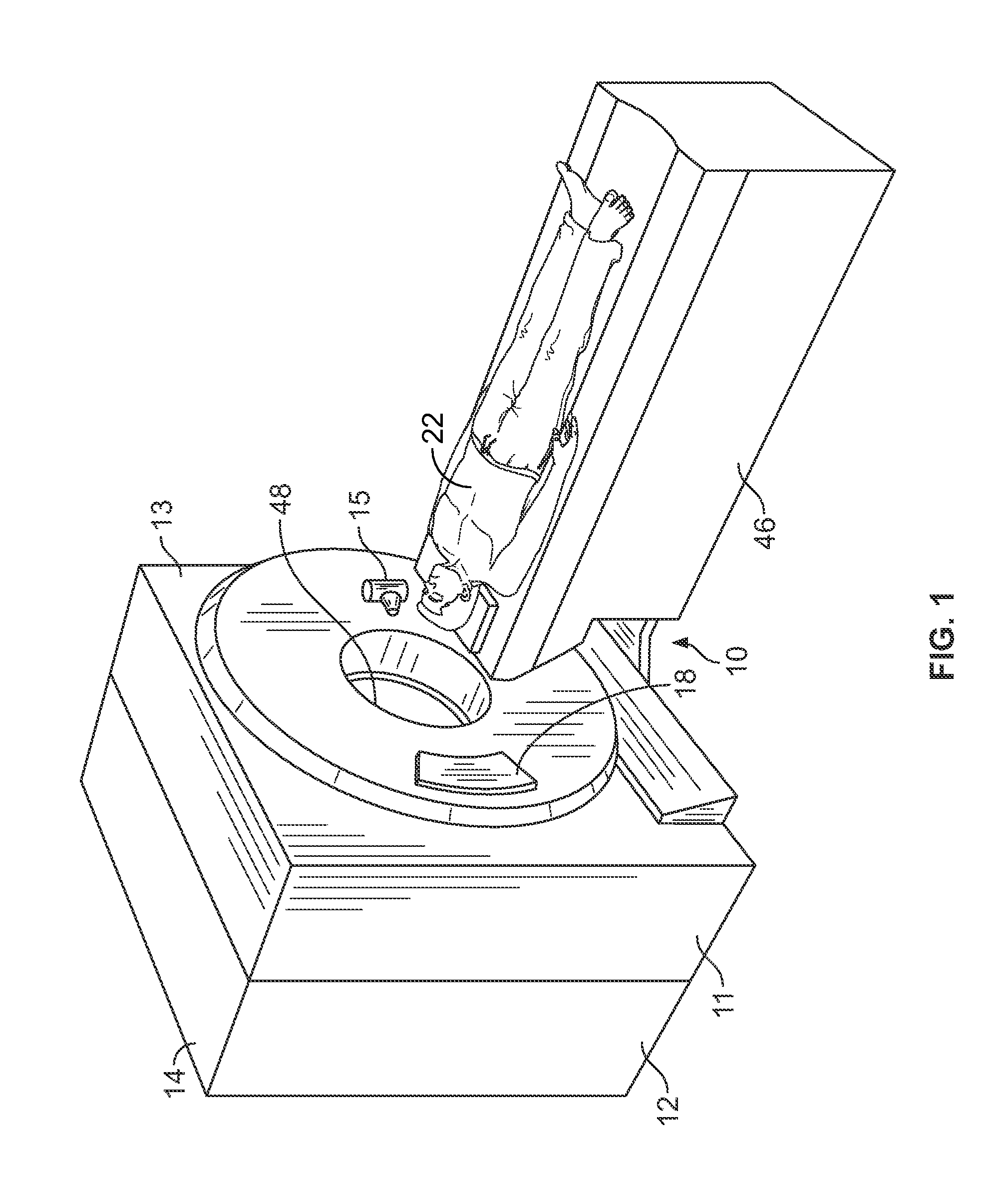
[0015]FIG. 1 is a pictorial view of an exemplary multi-modal imaging system 10 in accordance with an embodiment of the present invention. FIG. 2 is a block schematic diagram of the multi-modal imaging system 10 illustrated in FIG. 1 in accordance with an embodiment of the present invention. Although embodiments of the present invention are described in the context of an exemplary dual modality imaging system that includes a CT imaging system and a PET imaging system it should be understood that other imaging systems capable of performing the functions described herein are contemplated as being used.

[0016]There is herein provided a system and method for correcting multi-modality imaging data. The apparatus and methods are illustrated with reference to the figures wherein similar numbers indicate the same elements in all figures. Such figures are intended to be illustrative rather than limiting and are included herewith to facilitate explanation.

[0017]As discussed above, at least one ...


PUM


Abstract

A method for correcting Positron Emission Tomography (PET) data includes adjusting a tube current generated by the CT imaging system to a second tube current value that is less than a first tube current value used to generate diagnostic quality CT images, and imaging the patient with the CT imaging system set at the second tube current value. The method also includes generating a plurality of computed tomography (CT) projection data from the CT imaging system and preprocessing the CT projection data to generate preprocessed CT projection data. The method further includes filtering the preprocessed CT projection data to reduce electronic noise to generate filtered CT projection data, and performing a minus logarithmic operation on the filtered CT projection data to generate the corrected PET data.

Description

BACKGROUND OF THE INVENTION[0001]This invention relates generally to imaging systems capable of operation in multiple modalities, and more particularly to an apparatus and method for correcting multi-modality imaging data.[0002]Multi-modal imaging systems are capable of scanning using different modalities, such as, for example, but not limited to, Positron Emission Tomography (PET), Single photon emission computed tomography (SPECT), and Computed Tomography (CT). Conventional PET / CT imaging systems obtain CT images for attenuation correction in PET and to provide anatomical information to aid a physician in localizing tracer uptake used during PET imaging of a patient. In some cases, the CT diagnostic images are read separately. The accuracy of the reconstructed CT numbers is important to ensure adequate representation of the tracer uptake in the organ. Under normal scanning conditions, this constraint can be easily met by the CT imaging system because of the tight tolerances on the...

Claims


Application Information

Patent Timeline
Patent Type & Authority Patents(United States)
IPC IPC(8): G06K9/00
CPCA61B6/032A61B6/037A61B6/5235
Inventor HSIEH, JIANGCOLSHER, JAMES GEORGELONN, ALBERT HENRYGANIN, ALEXANDERTHIBAULT, JEAN-BAPTISTE
Owner GENERAL ELECTRIC CO
Learn More