
High tibial osteotomy system

a high-tibial osteotomy and tibial bone technology, applied in the field of high-tibial bone osteotomy system, can solve the problems of varus or valgus defect in the tibia, increased loading on the medial joint compartment, and increased loading on the lateral joint compartmen

Active Publication Date: 2012-08-14
HOWMEDICA OSTEONICS CORP
View PDF77 Cites 189 Cited by
  • Summary
  • Abstract
  • Description
  • Claims
  • Application Information

AI Technical Summary

Benefits of technology

The present invention relates to a cutting block for use in a bone osteotomy procedure. The cutting block has three cutting guide surfaces that can be affixed to the bone in two different positions, allowing for precise and safe cutting of the bone. The cutting block can be used in a high-tibial osteotomy procedure, where the first and second cutting guide surfaces are positioned on the bone's first side and the third cutting guide surface is positioned on the bone's second side. The cutting block can be easily attached and detached from the bone, and the three cutting guide surfaces form an intersecting angle to each other. The technical effects of the invention include improved accuracy and safety in bone osteotomy procedures and simplified and efficient procedures for preparing bone w wedges.

Problems solved by technology

Such uneven weight bearing results from either a varus or valgus defect in the tibia.
A varus deformity generally results in increased loading on the medial joint compartment, while a valgus defect results in increased loading on the lateral joint compartment.
Since the base length of the wedge spans the entire tibial medial-lateral width, the amount of distance to be closed is quite significant, resulting in shortening of the tibia, which may require corresponding shortening of the fibula.
It is problematic to shorten the length of the tibia because this can lead to problems in the gait of the patient leading to back, hip or other joint problems, and can complicate any subsequent total knee replacement (“TKR”) procedure which may be necessary.
Further, if shortening of the tibia requires that the fibula be shortened, the HTO procedure is further complicated and patient pain and recovery time are increased.
Additionally, tibial shortening leads to a significant amount of soft tissue laxity of the lateral compartment, which includes the joint capsule, lateral collateral ligament, etc.
Other complications arise from a closing wedge HTO procedure because the resected tibial plateau is “hinged” medially, for example.
This can result in the resected joint being extremely unstable during healing.
Subsequent TKR is further complicated by the presence of the staple used to secure the joint during healing because the staple may have to be removed prior to TKR.
Specifically, the amount of the distance to be “opened” can be quite significant, leading to lengthening of the leg and an undesirable amount of soft tissue tensioning of, for example, the medial compartment, which includes the joint capsule, the medial collateral ligament, etc.
Furthermore, the lateral or medial hinging of the osteotomy makes it extremely unstable, due to the amount of leverage applied to the hinge section from the opposite side of the tibia.
This instability makes the knee unable to resist torsional loads applied to the joint.
The presence of such hardware can complicate any subsequent TKR procedure which may be required.
Additionally, due to the large size of the wedge inserted into the cut, resorption and incorporation of a bone substitute device that may be used to fill the wedge is lengthy.
This harvesting requires a separate procedure to be performed to harvest the autograft, which adds time, pain, blood loss and the risk of infection to the procedure.
By taking either a medial or lateral approach for cutting, the patellar tendon is easily avoided; however, these approaches make alignment of cuts more difficult, because the mechanical axis is not visible from the side of the knee.
The total bisection of both the proximal tibia and fibula leads to an extremely unstable joint, which requires a great deal of hardware for stabilization, which would, most likely, need to be removed prior to subsequent TKR.
However, it is generally regarded as too invasive or risky for practical purposes.

Method used


Image

  • High tibial osteotomy system
  • High tibial osteotomy system
  • High tibial osteotomy system

Examples


Embodiment Construction

[0045]In describing the preferred embodiments of the subject matter illustrated and to be described with respect to the drawings, specific terminology will be resorted to for the sake of clarity. However, the invention is not intended to be limited to the specific terms so selected, and it is to be understood that each specific term includes all technical equivalents which operate in a similar manner to accomplish a similar purpose.

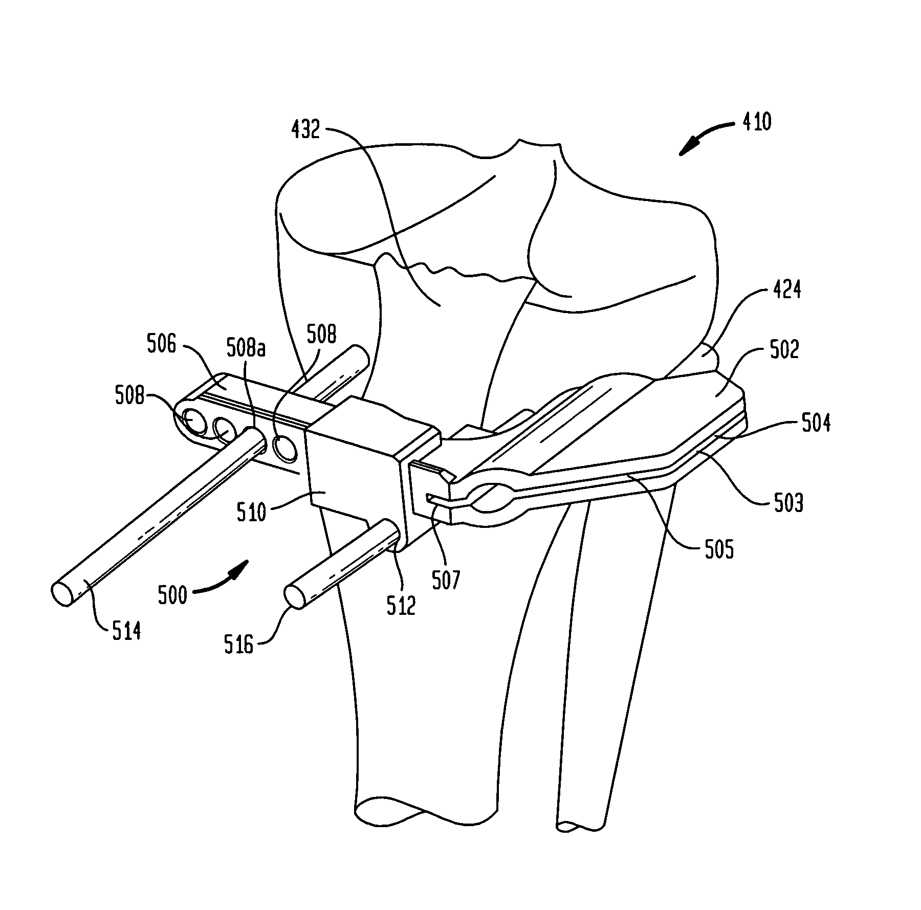
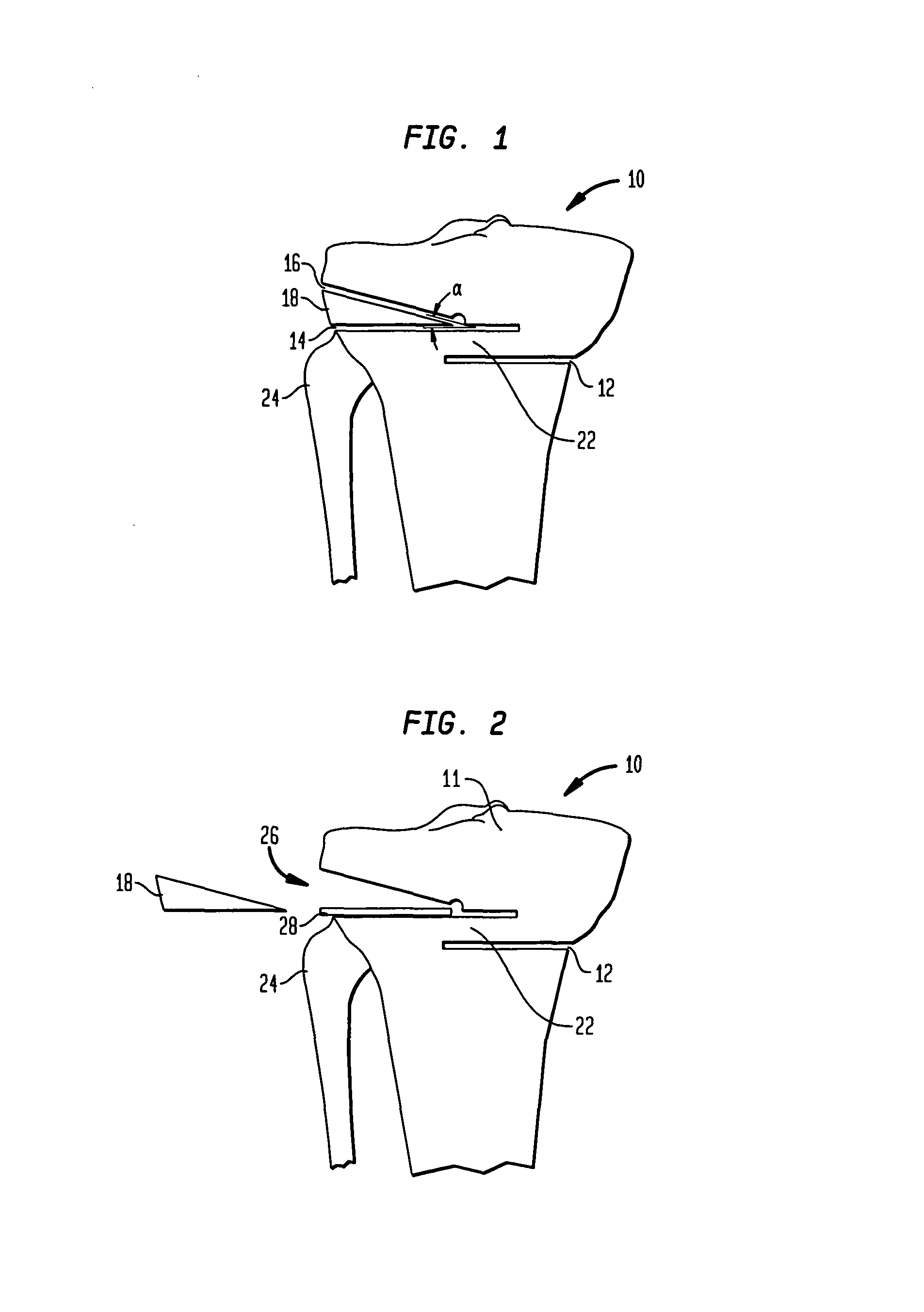
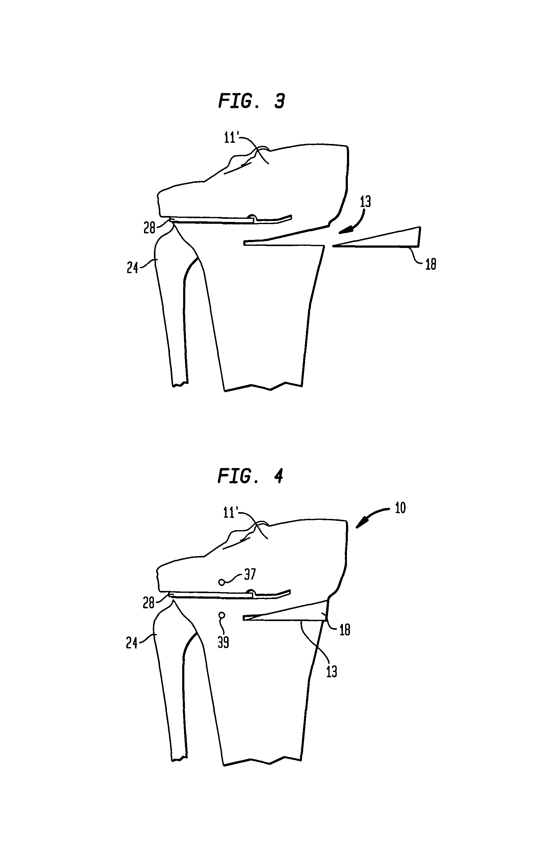
[0046]Referring to the drawings, wherein like reference numerals represent like elements, there is shown in FIGS. 1-4, in accordance with one embodiment of the present invention, an anterior view of the right tibia 10 with a series of cuts which can be used to complete an HTO procedure. The illustration in FIG. 1 shows exemplary locations for cuts in an HTO procedure used to correct a varus defect on the proximal tibia 11; however, as would be understood by one having reasonable skill in the art upon reading this disclosure, the procedure of the present i...


PUM


Abstract

A cutting block for use in a bone osteotomy procedure includes a first cutting guide surface, a second cutting guide surface, and a third cutting guide surface. The first, second, and third cutting guide surfaces are adapted to be temporarily affixed to a bone having a first side and a second side such that the first cutting guide surface is disposed on the first side of the bone, and such that the second cutting guide surface and third cutting guide surface are disposed on the second side of the bone forming an angle therebetween.

Description

CROSS-REFERENCE TO RELATED APPLICATIONS[0001]This application is related to U.S. application Ser. No. 11 / 478,790, filed on Jun. 30, 2006, and U.S. application Ser. No. 11 / 480,648, filed on Jul. 3, 2006, the disclosures of which are incorporated herein by reference.BACKGROUND OF THE INVENTION[0002]High tibial osteotomy (“HTO”) procedures have become well-established means of treating unicompartmental degenerative arthritis of the knee. This condition occurs due to uneven weight bearing of the femoral condyles on either medial or lateral joint compartments of the tibia. Such uneven weight bearing results from either a varus or valgus defect in the tibia. A varus or valgus defect occurs when the mechanical axis of the knee joint shifts either medially (valgus) or laterally (varus) of the preferred location therefor. It is generally accepted that the preferred location for the mechanical axis of the knee is at 62% of the tibial plateau from medial to lateral. The process for determining...

Claims


Application Information

Patent Timeline
Patent Type & Authority Patents(United States)
IPC IPC(8): A61B17/56
CPCA61B17/152A61B17/1604A61B17/1675A61B2017/565A61F2002/2892A61F2002/30736A61F2002/30892A61B17/8095A61B17/157
Inventor COLLAZO, CARLOS E.
Owner HOWMEDICA OSTEONICS CORP
Learn More