
Internal prosthesis for reconstruction of cardiac geometry

a technology of internal prosthesis and cardiac geometry, which is applied in the field of heart surgery and the treatment of congestive heart failure, can solve the problems of cardiac dyspnea, cardiac dyspnea, chest pain, and other adverse clinical symptoms, and achieve the effect of simple and easy-to-use cardiac prosthesis

Inactive Publication Date: 2012-06-26
THE INT HEART INST OF MONTANA FOUND
View PDF36 Cites 211 Cited by
  • Summary
  • Abstract
  • Description
  • Claims
  • Application Information

AI Technical Summary

Benefits of technology

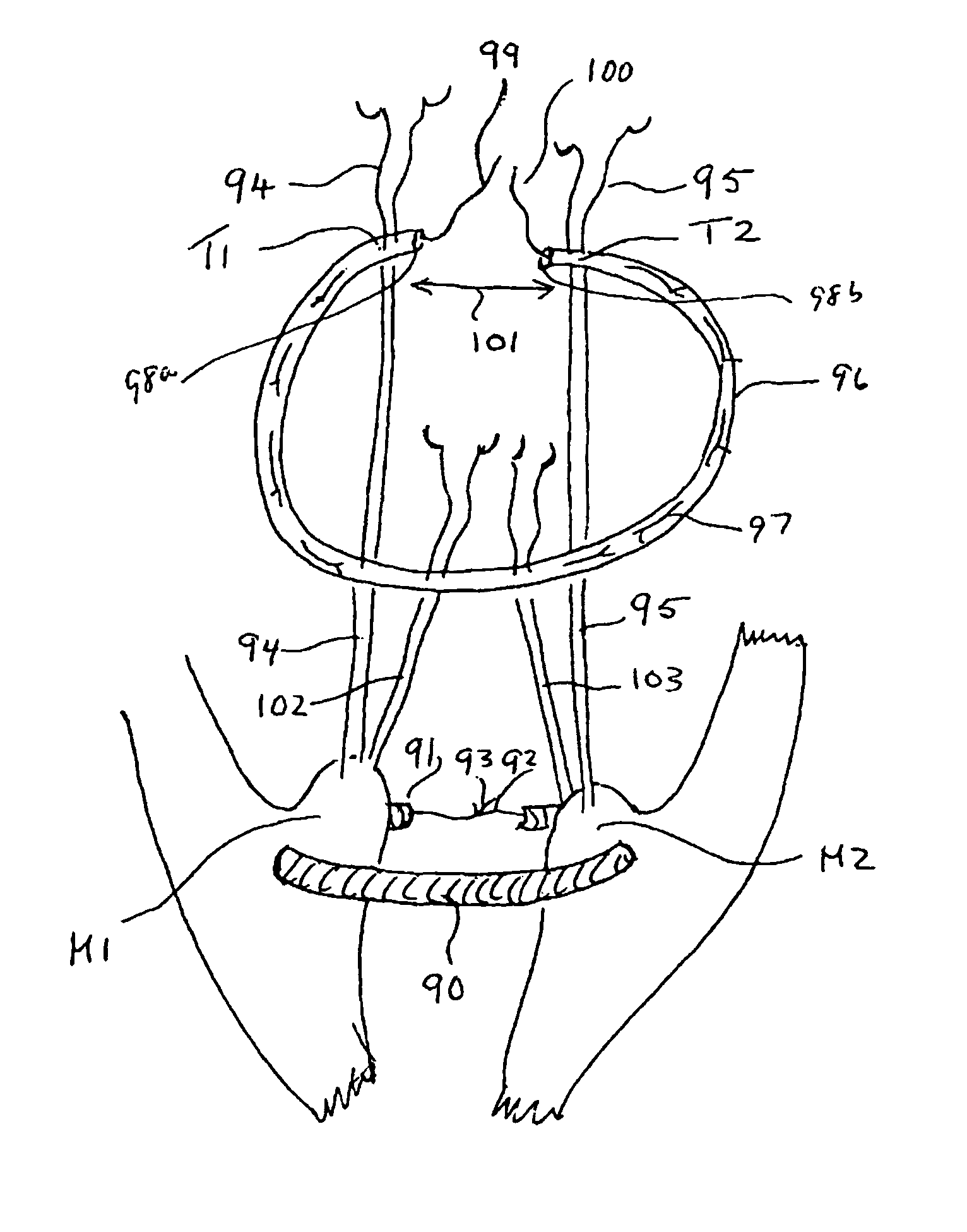
[0010]The present invention provides a unique unexpectedly simple and easy to use cardiac prosthesis that includes a semi-circular band portion having first and second ends. The first end includes a first portion of a tether and the second end includes a second portion of the tether, such that when the first and second portions of the tether are secured to each other, the ultimate assembly provides an adjustable semi-circular band. This band can be affixed to an annulus of a valve, or can be inserted through trabeculae associated with papillary muscles.
[0016]The method of the invention can further include securing synthetic or biologic chords from the papillary muscle to the annuloplasty prosthesis. There are several ways to accomplish this. The chords can be inserted through tissue about each prosthesis. Additional securing can be accomplished by use of pledgets to help prevent damage to the tissue. Alternatively, the replacement chords can be inserted through each prosthesis, thereby minimizing potential damage to the surrounding tissue. The chords can be made of materials known in the art, such as nylons, polypropylene, polyesters, polyurethanes and the like or made of biologic membranes. The replacement chord can also be a rigid rod. In any event, the synthetic chords will have a length wherein the relationship between the lengths of the chords are adjusted such that the distance from the tips of the papillary muscles is approximately equal to the intertrigonal distance of the valve annulus.
[0018]To help estimate the intertrigonal distance and assist with the methods of the invention, the present invention also provides a trigonal-papillary sizing device. The trigonal-papillary sizing device includes a handle attached to a two forked prong. The two forked prong includes a proximal portion and two distal ends each having an equivalent length, wherein the proximal portion connects the two prongs. The two distal prong ends are generally rounded or configured in such a fashion as to not damage tissue as the device is being manipulated in the ventricular chamber. The distance between the two prongs and the lengths of the two prongs are equivalent, the distances being equal to the intertrigonal distance of the valve. Sizing devices can be constructed such that the operator can quickly choose the appropriate size by comparing the distance between the forks with the intertrigonal distance. The present invention, therefore provides, intertrigonal-papillary sizing devices of varying dimensions that are suited to various sized hearts.
[0020]The present invention also provides methods to align the papillary muscles of the ventricle. This is accomplished by forming a passage about the papillary muscles of the ventricle through the surrounding trabeculae. The cardiac prosthesis of the invention, as described throughout the specification, is guided through and adjusted via the tether, such that the papillary muscles and chords are aligned relative to the mitral valve annulus. In an alternative embodiment, the cardiac prosthetic can be guided through the trabeculae simultaneously while forming the passage, or the device can be configured such that a needle or hook is attached to either end of the tether and then removed prior to securing the tether. Optionally a biocompatible tape, such as Gore-Tex could be used. Additionally, pledgets can be secured to the papillary muscles with the suture or tape to prevent damage to the papillary tissue.
[0024]The method can further include securing chords from the cardiac prosthesis to the mitral valve annuloplasty prosthesis. There are several ways to accomplish this. The chords can be inserted through tissue about each prosthesis. Additional securing can be accomplished by use of pledgets to help prevent damage to the tissue. Alternatively, the chords can be inserted through each prosthesis, thereby minimizing potential damage to the surrounding tissue. The chords can be made of materials known in the art, such as nylons, polypropylene, polyesters, polyurethanes and the like or biologic membrane. The chord can also be a rigid rod. In any event, the chords will have a length wherein the relationship between the lengths of the chords are adjusted such that the distance from the tips of the papillary muscles is equal to the intertrigonal distance of the mitral valve annulus.

Problems solved by technology

The result of valve regurgitation is often associated with arrhythmias, chest pain, cardiac dyspnea, and other adverse clinical symptoms.
This distortion of the valve geometry increases the mechanical stress of the myocardial fibers initiating a downward spiral of the ventricular contractility.

Method used


Image

  • Internal prosthesis for reconstruction of cardiac geometry
  • Internal prosthesis for reconstruction of cardiac geometry
  • Internal prosthesis for reconstruction of cardiac geometry

Examples


Embodiment Construction

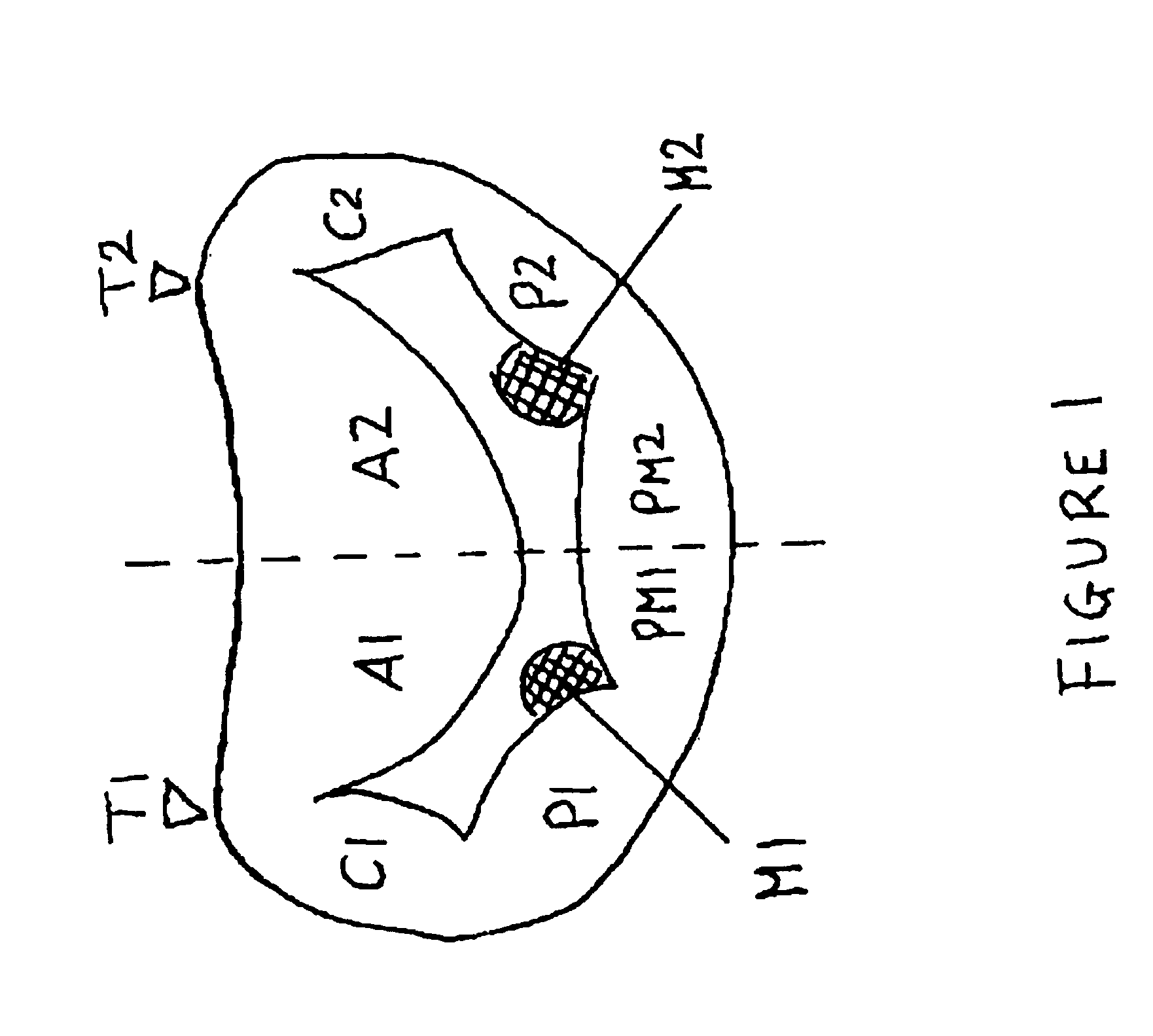
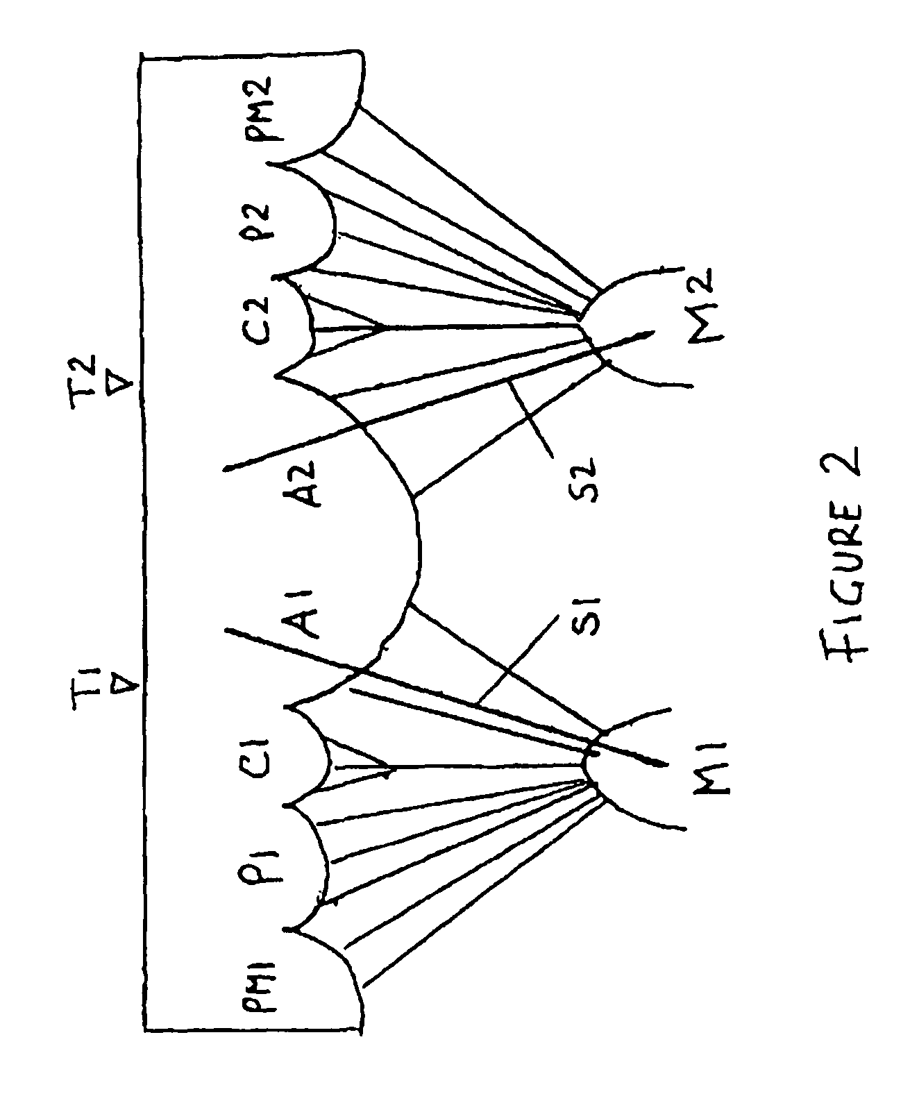
[0043]In congestive heart failure the dimensions of the mitral apparatus are severely altered. 1) The whole mitral annulus is dilated but non homogeneously. This dilatation includes the intertrigonal distance and posterior annulus but is particularly severe in its antero-posterior diameter. This dilatation reduces the leaflet coaptation generating mitral regurgitation. 2) The papillary muscles are displaced laterally and apically tethering the leaflets and consequently reducing their mobility and increasing the valve regurgitation. 3) The papillary displacement also pulls on the basal stay chords deforming the anterior leaflet.

[0044]The present invention, therefore, provides for the complete internal reconstruction of the mitral apparatus and includes a mitral annuloplasty, a papillary plasty and the implantation of new stay chords that brings closer the papillary muscles to the trigones of the mitral annulus. These aims require not only suitable devices but also guidelines easy to ...


PUM


Abstract

Unique semi-circular papillary muscle and annulus bands are described that are useful for modifying the alignment of papillary muscles, a mitral valve annulus and / or a tricuspid valve annulus. Methods to effect the alignment are also described and an unique sizing device that can be used for such alignment is described.

Description

CROSS REFERENCE TO RELATED APPLICATION[0001]This application claims benefit under 35 U.S.C. §119(e) to application Ser. No. 60 / 546,872, entitled “Novel System for Restoration of the Failing Heart” by Carlos, M. G. Duran on Feb. 23, 2004, the contents of which are incorporated herein in their entirety.FIELD OF THE INVENTION[0002]The present invention relates to heart surgery and to the treatment of congestive heart failure.BACKGROUND OF THE INVENTION[0003]The heart has four chambers, the left and right atria and the left and right ventricles. The atria collect blood as it returns from the body in the case of the right atrium or from the lungs in the case of the left atrium. During diastole the atrioventricular valves (tricuspid valve on the right side and mitral on the left) open, filling the ventricles. During systole the ventricles contract closing the atrioventricular valves and expelling the blood towards either the body (left) or the lungs (right).[0004]The bicuspid or mitral va...

Claims


Application Information

Patent Timeline
Patent Type & Authority Patents(United States)
IPC IPC(8): A61F2/24
CPCA61F2/2448A61F2/2496A61F2/2457
Inventor GOMEZ DURAN, CARLOS MANUEL
Owner THE INT HEART INST OF MONTANA FOUND
Learn More