
Method and apparatus for the non-invasive measurement of tissue function and metabolism by determination of steady-state fluorescence anisotropy

a steady-state fluorescence anisotropy and tissue function technology, applied in the field of biological tissue non-invasive measurement of functions, can solve the problems of crude resolution, high implementation cost, and inability to accurately measure the so as to achieve calibration-free measurement of metabolic rate and function, non-invasive determination, and high resolution. the effect of high accuracy

Inactive Publication Date: 2012-03-06
MOORING SYST
View PDF22 Cites 25 Cited by
  • Summary
  • Abstract
  • Description
  • Claims
  • Application Information

AI Technical Summary

Benefits of technology

This approach allows for high-resolution, non-invasive, and sensitive measurement of tissue metabolism and function in 2- and 3-dimensional spaces, enabling early disease detection and prognosis before irreversible structural damage occurs, while avoiding tissue damage from high-energy pulses.

Problems solved by technology

Structural damage in living tissues is always accompanied by functional deficit.
However, the converse is not necessarily true.
PET scanning provides striking images of functional change; however, its resolution is crude and its implementation expensive.
However, these approaches have never successfully been extended to the clinical setting because they require calibration of the system by bringing the tissue to a uniform oxygen partial pressure of 0 mm Hg by breathing an animal on 100% nitrogen.
This obviously cannot be done in humans without causing irreversible cell death.
To overcome these deficiencies, some have tried to take the ratio of emissions from NADH and FAD, but the results have not been satisfactory because the rates of photobleaching of these molecules are different, thus precluding a clinically useful tool.
However, such intensity-based methods are severely limited due to photobleaching, inner filter effects and the difficulties associated with isolating contributions from these metabolically relevant fluorophores from the background autofluorescence of other endogenous fluorophores.
Although measurements of fluorescence lifetime can, to some extent, overcome the limitations of intensity-based measurements, the information gleaned from such measurements alone is limited, and ultra fast lifetimes require the use of high energy laser pulses that may be damaging to fragile tissues.
Moreover, fluorescence anisotropy and lifetime are intrinsic parameters, unlike the intensity signals used to compute them, and are therefore insensitive to light path and geometry.
It should also be noted that time-resolved fluorescence anisotropy measurement requires the use of ultra-fast laser pulses that would similarly be destructive to tissues.

Method used


Image

  • Method and apparatus for the non-invasive measurement of tissue function and metabolism by determination of steady-state fluorescence anisotropy
  • Method and apparatus for the non-invasive measurement of tissue function and metabolism by determination of steady-state fluorescence anisotropy
  • Method and apparatus for the non-invasive measurement of tissue function and metabolism by determination of steady-state fluorescence anisotropy

Examples


Embodiment Construction

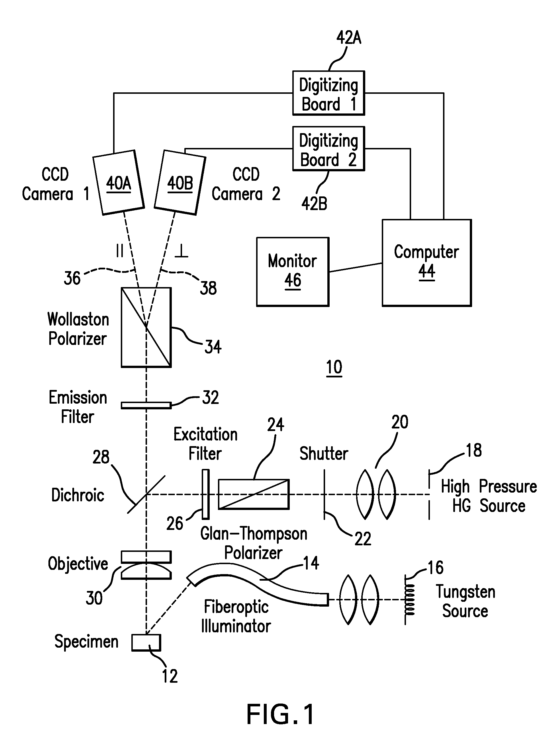
[0045]FIG. 1 provides a schematic diagram of an apparatus 10 designed for the topographic, 2-dimensional, mapping of tissue function and metabolism non-invasively in an imaged tissue. Specimen 12 includes one or more biological tissues which contain one or more substances, called fluorophors, which fluoresce when illuminated.

[0046]Specimen 12 is illuminated with nonpolarized visible light by a fiberoptic illuminator 14 utilizing a tungsten source 16. The radiant energy of a xenon arc lamp 18 is gathered by a collector lens 20, and is selectively allowed to pass through shutter 22. The light then passes through a Glan-Thompson polarizer 24 (obtained from Ealing, Inc.).

[0047]The light is spectrally shaped by an excitation filter 26 and is then reflected by a dichroic mirror 28 (obtained from Omega Optical) which reflects excitation wavelengths through an objective lens 30 to the imaged tissue, causing fluorophors in the tissue to fluoresce.

[0048]Emitted luminescence from the excited t...


PUM


Abstract

A non-invasive measurement of biological tissue reveals information about the function of that tissue. Polarized light is directed onto the tissue, stimulating the emission of fluorescence, due to one or more endogenous fluorophors in the tissue. Fluorescence anisotropy is then calculated. Such measurements of fluorescence anisotropy are then used to assess the functional status of the tissue, and to identify the existence and severity of disease states. Such assessment can be made by comparing a fluorescence anisotropy profile with a known profile of a control.

Description

CROSS-REFERENCE TO PRIOR APPLICATION[0001]Priority is claimed from U.S. Provisional Patent Application Ser. No. 60 / 744,831, filed Apr. 13, 2006, the entire disclosure of which is incorporated by reference herein.BACKGROUND OF THE INVENTION[0002]This invention relates to the field of non-invasive measurements of functions of biological tissues.[0003]Structural damage in living tissues is always accompanied by functional deficit. However, the converse is not necessarily true. That is, functional deficit may precede irreversible structural damage for many years, in numerous disease states, and may therefore serve as a diagnostic indicator of early disease and a prognostic indicator of disease progression. For this reason, increasing attention has focused on functional imaging of tissues, in situ, in humans.[0004]PET scanning provides striking images of functional change; however, its resolution is crude and its implementation expensive.[0005]To provide high-resolution imaging of the fu...

Claims


Application Information

Patent Timeline
Patent Type & Authority Patents(United States)
IPC IPC(8): C12Q1/00
CPCA61B5/14546A61B5/1455A61B5/14555A61B5/14556G01N21/645A61B5/0071G01N21/6445G01N2021/6484A61B5/4842A61B5/7246A61B5/7278G01N2201/0683G01N2201/12
Inventor ZUCKERMAN, RALPH
Owner MOORING SYST
Learn More