
Method and apparatus for detecting arrhythmias in a subcutaneous medical device

a medical device and arrhythmia detection technology, applied in the field of implantable medical devices, can solve the problems of inability to place endocardial leads, risk of future sudden death episodes, and general recommendation not recommended for pediatric cardiac patients

Active Publication Date: 2011-08-02
MEDTRONIC INC
View PDF48 Cites 143 Cited by
  • Summary
  • Abstract
  • Description
  • Claims
  • Application Information

AI Technical Summary

Benefits of technology

The present invention relates to an improved method and apparatus for detecting arrhythmias in a subcutaneous implantable medical device. The technical effects of the invention include reliably sensing and detecting arrhythmias while rejecting noise and other physiologic signals, as well as improving the accuracy of detecting arrhythmias in patients who may be at risk of sudden cardiac death. The invention also addresses the challenge of limited sensing in a subcutaneous device due to the absence of intracardial or epicardial electrodes. Additionally, the invention addresses the potential for inappropriate therapy by the subcutaneous device when combined with an existing pacemaker in a patient.

Problems solved by technology

Attempts are being made to identify patients who are asymptomatic by conventional measures but are nevertheless at risk of a future sudden death episode.
Further, endocardial lead placement is not possible with patients who have a mechanical heart valve implant and is not generally recommended for pediatric cardiac patients.
There are technical challenges associated with the operation of a SubQ device.
This is particularly because the SubQ device is limited to far-field sensing since there are no intracardial or epicardial electrodes in a subcutaneous system.
Further, sensing of atrial activation from subcutaneous electrodes is limited since the atria represent a small muscle mass and the atrial signals are not sufficiently detectable transthoracically.
Yet another challenge could occur in situations where it is desirable to combine a SubQ device with an existing pacemaker (IPG) in a patient.
While this may be desirable in a case where an IPG patient may need a defibrillator, a combination implant of a SubQ device and an IPG may result in inappropriate therapy by the SubQ device, which may pace or shock based on spikes from the IPG.
The result can be over-counting beats from the atrium, ventricles or both; or, because of the larger pacing spikes, sensing of arrhythmic signals (which are typically much smaller in amplitude) may be compromised.

Method used


Image

  • Method and apparatus for detecting arrhythmias in a subcutaneous medical device
  • Method and apparatus for detecting arrhythmias in a subcutaneous medical device
  • Method and apparatus for detecting arrhythmias in a subcutaneous medical device

Examples


Embodiment Construction

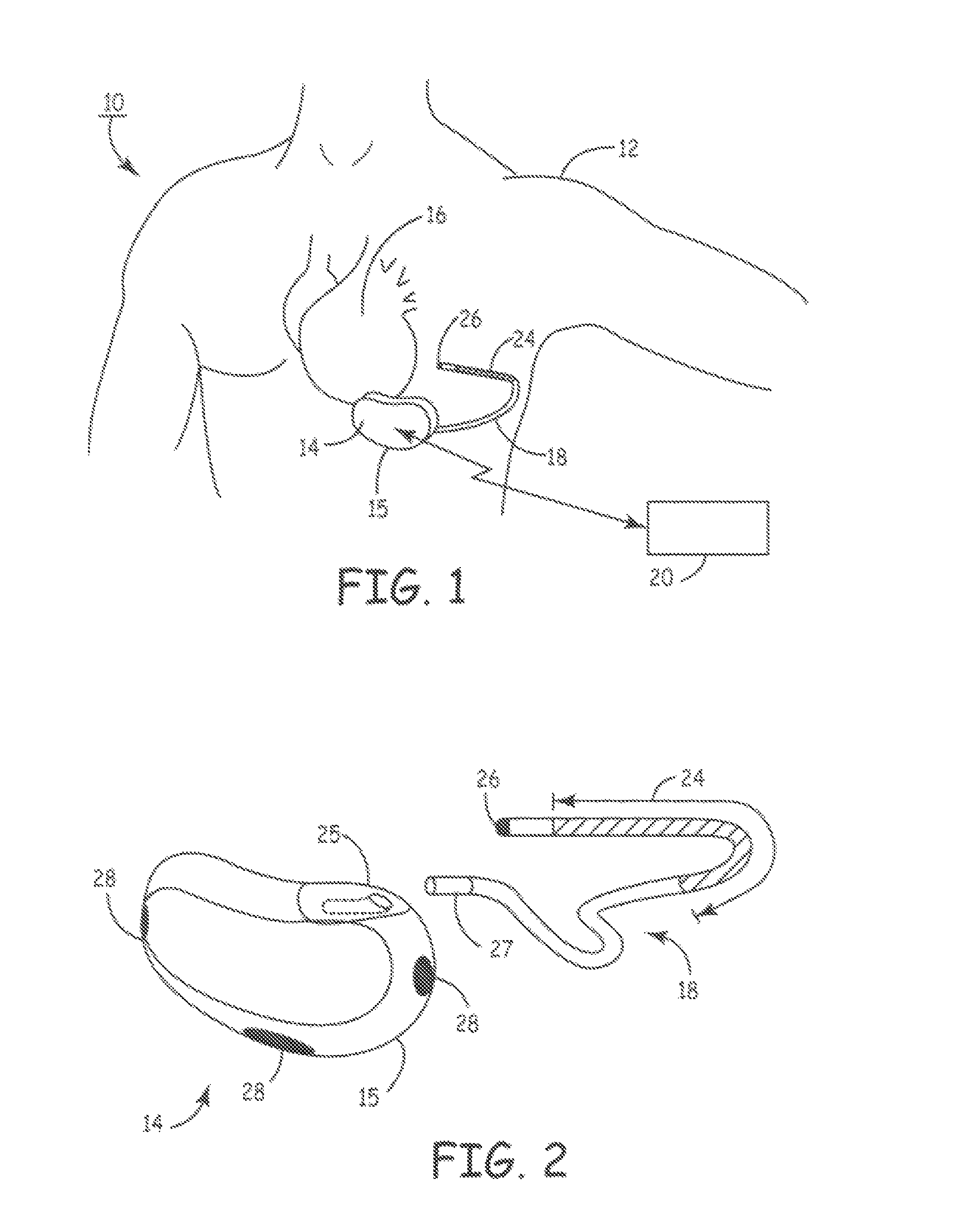
[0025]FIG. 1 is a schematic diagram of an exemplary subcutaneous device in which the present invention may be usefully practiced. As illustrated in FIG. 1, a subcutaneous device 14 according to an embodiment of the present invention is subcutaneously implanted outside the ribcage of a patient 12, anterior to the cardiac notch. Further, a subcutaneous sensing and cardioversion / defibrillation therapy delivery lead 18 in electrical communication with subcutaneous device 14 is tunneled subcutaneously into a location adjacent to a portion of a latissimus dorsi muscle of patient 12. Specifically, lead 18 is tunneled subcutaneously from the median implant pocket of the subcutaneous device 14 laterally and posterially to the patient's back to a location opposite the heart such that the heart 16 is disposed between the subcutaneous device 14 and the distal electrode coil 24 and distal sensing electrode 26 of lead 18.

[0026]It is understood that while the subcutaneous device 14 is shown positi...


PUM


Abstract

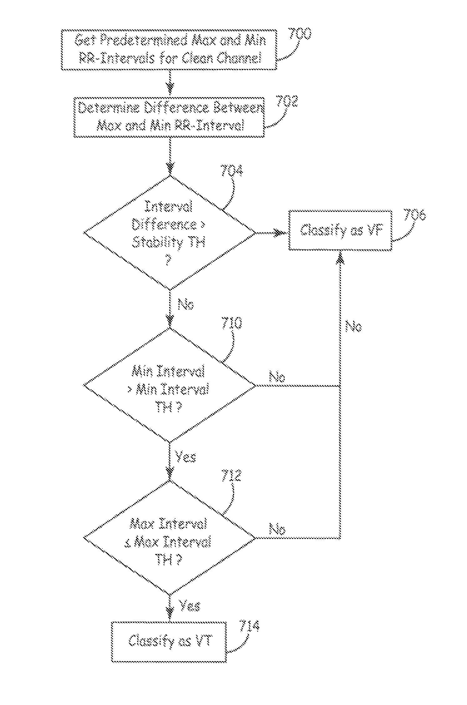
A method and apparatus for detecting a cardiac event in a medical device that includes sensing cardiac signals from a plurality of electrodes forming a first sensing vector and a second sensing vector, advancing from a first state to a second state in response to processing of an interval associated with a cardiac cycle of the sensed cardiac signals, and advancing from the second state in response to processing of a cardiac signal sensed during a predetermined sensing window.

Description

RELATED APPLICATION[0001]The present application claims priority and other benefits from U.S. Provisional Patent Application Ser. No. 60 / 786,981, filed Mar. 29, 2006, entitled “METHOD AND APPARATUS FOR DETECTING ARRHYTHMIAS IN A SUBCUTANEOUS MEDICAL DEVICE”, incorporated herein by reference in its entirety.FIELD OF THE INVENTION[0002]The present invention generally relates to an implantable medical device system, and more particularly to a method and apparatus for detecting arrhythmias in a subcutaneous medical device.BACKGROUND OF THE INVENTION[0003]Many types of implantable medical devices (IMDs) have been implanted that deliver relatively high-energy cardioversion and / or defibrillation shocks to a patient's heart when a malignant tachyarrhythmia, e.g., ventricular tachycardia or ventricular fibrillation, is detected. Cardioversion shocks are typically delivered in synchrony with a detected R-wave when fibrillation detection criteria are met, whereas defibrillation shocks are typi...

Claims


Application Information

Patent Timeline
Patent Type & Authority Patents(United States)
IPC IPC(8): A61N1/368A61B5/361A61B5/363
CPCA61N1/3621A61N1/3956A61B5/046A61B5/0464A61B5/7239A61B5/361A61B5/363
Inventor GHANEM, RAJA N.STADLER, ROBERT W.ZHANG, XUSHENG
Owner MEDTRONIC INC
Learn More