
Method and system to treat and prevent myocardial infarct expansion

a myocardial infarct and systemic technology, applied in the field of myocardial infarct expansion treatment and system, can solve the problems of reducing cardiac output, reducing cardiac output, and reducing structure collapse, so as to reduce the severity and complications of mi, reduce the stress of the infarcted ventricle, and reduce the severity of the infarct expansion

Inactive Publication Date: 2011-02-08
G&L CONSULTING
View PDF70 Cites 4 Cited by
  • Summary
  • Abstract
  • Description
  • Claims
  • Application Information

AI Technical Summary

Benefits of technology

[0040]In one embodiment, the invention reduces the severity and complications of MI by reducing the infarct expansion which may be achieved by reducing stress in the infarcted ventricle by constraining the heart and reducing the diastolic dilation of the heart. The infarct expansion may be reduced with a procedure that is practical, simple, easily reversible, and minimally invasive (does not require general anesthesia and surgery).
[0041]Multiple animal studies have established benefit of constraining the heart in hours and days immediately following MI. The purpose of a cardiac constraint is the reduction of the myocardial stress and ventricular dilation.
[0043]A catheter may be place in the pericardial space by performing a minimally invasive surgical procedure. For example, a 2 inch long incision may be made just below the last rib. Though the incision, the pericardium may be easily visualized. A small incision may be made in the pericardium through which the catheter may be placed into the pericardial space.
[0044]The pericardial sac can also be accessed with a percutaneous catheter therefore eliminating the need for surgery. Pressure in the pericardial sac can be controlled to ensure that the patient is not in danger. In case of emergency the fluid infused into the pericardial sac can be instantly drained therefore totally reversing the therapy.
[0047]Drainage of pericardial fluid called pericardiocentesis is the definitive treatment of cardiac tamponade. Removal of even a small volume of fluid can rapidly improve blood pressure and cardiac output, and may ultimately prove to be lifesaving. The pericardium can be tapped from almost any reasonable location on the chest wall. However, for the usual “blind” pericardiocentesis, the subxiphoid approach is preferred. Echocardiography is often used to guide needle insertion and the subsequent path of the needle / catheter. The patient may either be recumbent or positioned so that his chest is at a 30-degree angle from the bed. The needle is then inserted into the left xiphocostal angle perpendicular to the skin and 3 to 4 mm below the left costal margin. The needle is advanced directly into the inner aspect of the rib cage; the depth of perpendicular penetration depends on the amount of overlying soft tissue. The hub of the needle is then depressed so that the needle points toward the left shoulder. Using a slow, cautious “warm gear” turning action of the fingers, the needle is advanced about 5 to 10 mm of until fluid is reached.

Problems solved by technology

If the pressure exceeds atrial or ventricular diastolic (also called filling) pressure, these structures may collapse.
Collapse of the atria or ventricles will cause a limitation in filling of the heart, thus a decrease cardiac output.
Decreased cardiac output leading to hypotension is primarily responsible for causing death in the patients.

Method used


Image

  • Method and system to treat and prevent myocardial infarct expansion
  • Method and system to treat and prevent myocardial infarct expansion
  • Method and system to treat and prevent myocardial infarct expansion

Examples


Embodiment Construction

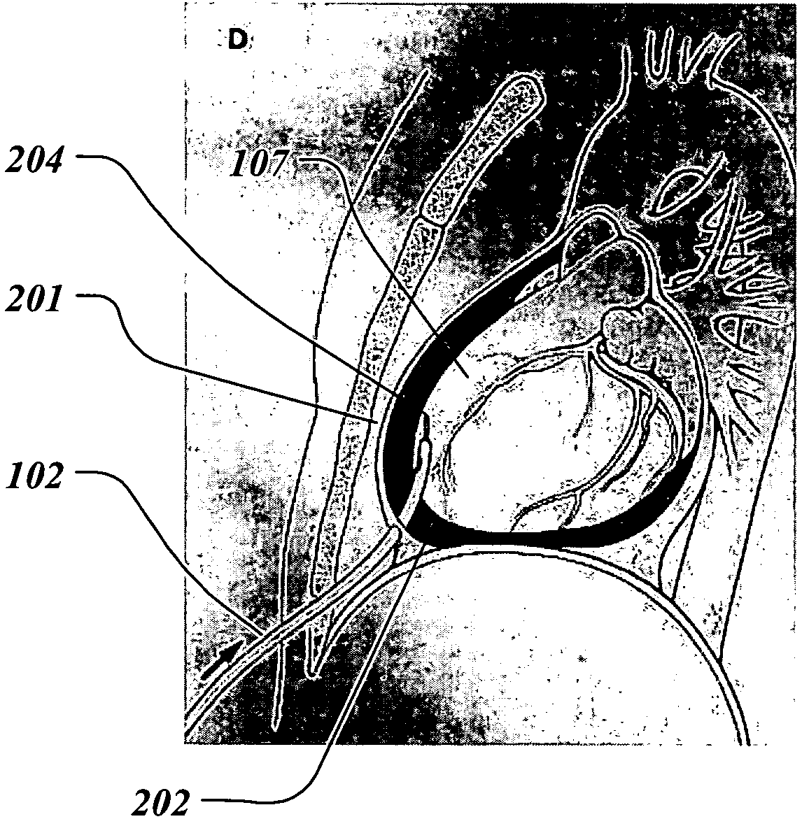
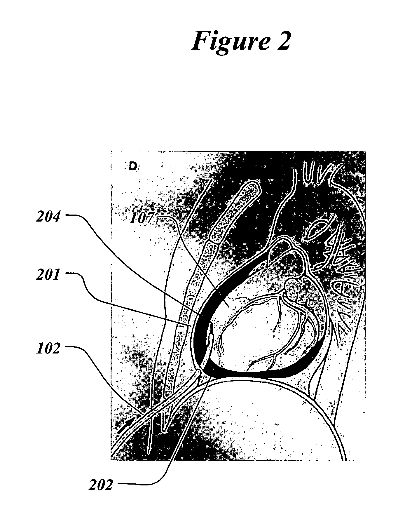
[0056]For the proposed clinical use, the capability of the preferred embodiment of the invention is to constrain the heart by controllably elevating hydraulic pressure in the pericardial sac.

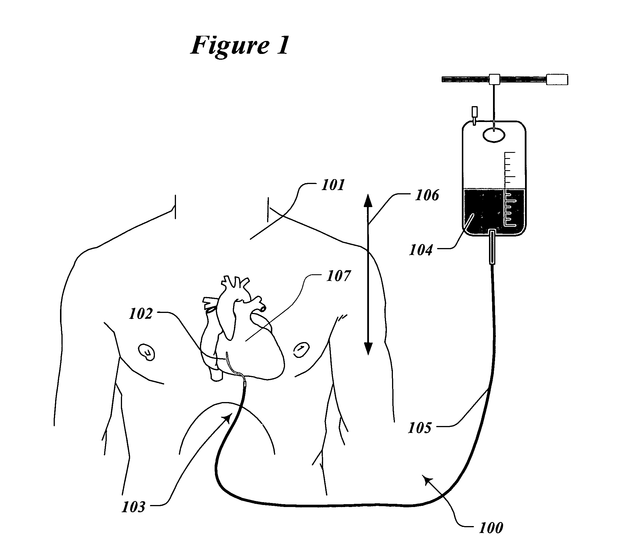
[0057]FIG. 1 illustrates the treatment of a patient 101 with the system 100 for infusion of fluid into the pericardial sac of the heart. The infusion catheter 102 is partially inserted into the pericardial sac of the heart 107. Catheter 102 crosses the patient's skin in the xiphoid area 103. The distal tip of the catheter 102 has an opening and is in fluid communication with the pericardial (also called intrapericardial) space. The proximal end of the catheter is connected to the bag 104 filled with the hydraulic infusion solution such as sterile saline by the fluid filled tube 105. The height difference 106 between the patient's heart 107 and the level of fluid in the bag determines the hydraulic pressure in the pericardium. For example, if the hydraulic fluid has the specific gravity of water,...


PUM


Abstract

A method and apparatus for prevention and reduction of myocardial infarct expansion and heart remodeling by infusion of fluid into the pericardial space of the heart to created a hydraulic heart constraint. As a result of the constraint ventricular stress and dilation is reduced. Pressure in the pericardial sac is maintained at a safe level for the duration of treatment. Apparatus consists of a catheter and a fluid infusion system.

Description

RELATED APPLICATION[0001]This applications claims the benefit of the U.S. Provisional application 60 / 457,246, filed Mar. 26, 2003, the entirety of which is incorporated by reference.FIELD OF THE INVENTION[0002]This invention relates to a method for preventing expansion of the myocardial infarct size following a heart attack. It also relates to the reduction or constraining of the dilation of the heart following an infarct. It also relates to pressure-controlled infusion of fluid into the pericardial space of the heart.BACKGROUND OF THE INVENTION[0003]A Myocardial Infarction (MI) or heart attack, occurs when the blood supply to some part of the heart muscle (myocardium) is abruptly stopped. This is often due to clotting in a coronary blood vessel. Blood supplying the heart muscle comes entirely from two coronary arteries, both lying along the outside surface of the heart. If one of these arteries or any part of one suddenly becomes blocked, the area of the heart being supplied by the...

Claims


Application Information

Patent Timeline
Patent Type & Authority Patents(United States)
IPC IPC(8): A61M31/00A61F2/958A61M25/02
CPCA61M25/02A61M25/1011A61M2025/0233A61M2210/122
Inventor LEVIN, HOWARD R.GELFAND, MARK
Owner G&L CONSULTING
Learn More